Forconi Catherine S, Oduor Cliff I, Oluoch Peter O, Ong'echa John M, Münz Christian, Bailey Jeffrey A, Moormann Ann M
Division of Infectious Diseases, Department of Medicine, University of Massachusetts, Worcester, MA, United States.
Department of Pathology and Laboratory Medicine, Warren Alpert Medical School, Brown University, Providence, RI, United States.
Front Cell Infect Microbiol. 2020 Apr 21;10:162. doi: 10.3389/fcimb.2020.00162. eCollection 2020.
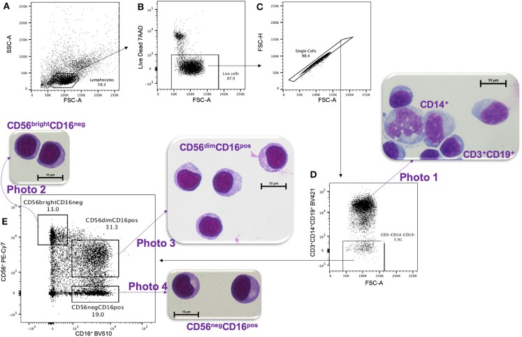
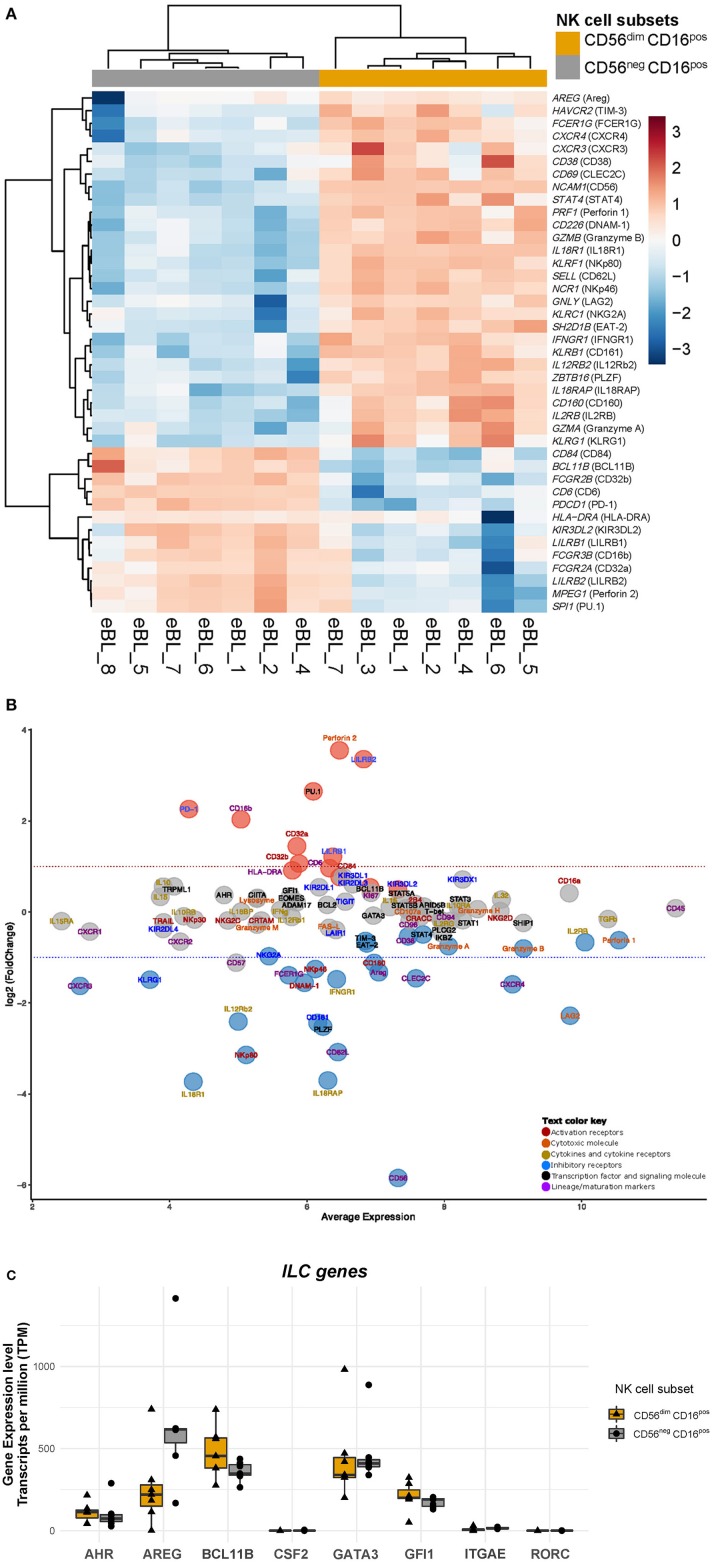
Natural Killer (NK) cells play an essential role in antiviral and anti-tumoral immune responses. In peripheral blood, NK cells are commonly classified into two major subsets: CD56CD16 and CD56CD16 despite the characterization of a CD56CD16 subset 25 years ago. Since then, several studies have described the prevalence of an CD56CD16 NK cell subset in viral non-controllers as the basis for their NK cell dysfunction. However, the mechanistic basis for their cytotoxic impairment is unclear. Recently, using a strict flow cytometry gating strategy to exclude monocytes, we reported an accumulation of CD56CD16 NK cells in malaria-exposed children and pediatric cancer patients diagnosed with endemic Burkitt lymphoma (eBL). Here, we use live-sorted cells, histological staining, bulk RNA-sequencing and flow cytometry to confirm that this CD56CD16 NK cell subset has the same morphological features as the other NK cell subsets and a similar transcriptional profile compared to CD56CD16 NK cells with only 120 genes differentially expressed (fold change of 1.5, < 0.01 and FDR<0.05) out of 9235 transcripts. CD56CD16 NK cells have a distinct profile with significantly higher expression of (perforin 2), (CD16b), , and (CD32A and B) as well as , and (PD-1), whereas Interleukin 18 (IL18) receptor genes ( and ), cytotoxic genes such as (NKp80) and (NKp46), and inhibitory (TIM-3) are significantly down-regulated compared to CD56CD16 NK cells. Together, these data confirm that CD56CD16 cells are legitimate NK cells, yet their transcriptional and protein expression profiles suggest their cytotoxic potential is mediated by pathways reliant on antibodies such as antibody-dependent cell cytotoxicity (ADCC), antibody-dependent respiratory burst (ADRB), and enhanced by complement receptor 3 (CR3) and FAS/FASL interaction. Our findings support the premise that chronic diseases induce NK cell modifications that circumvent proinflammatory mediators involved in direct cytotoxicity. Therefore, individuals with such altered NK cell profiles may respond differently to NK-mediated immunotherapies, infections or vaccines depending on which cytotoxic mechanisms are being engaged.
自然杀伤(NK)细胞在抗病毒和抗肿瘤免疫反应中发挥着重要作用。在外周血中,尽管25年前就已鉴定出CD56dimCD16bright亚群,但NK细胞通常被分为两个主要亚群:CD56brightCD16dim和CD56dimCD16bright。从那时起,多项研究描述了病毒无控制者中CD56dimCD16bright NK细胞亚群的普遍性,作为其NK细胞功能障碍的基础。然而,其细胞毒性受损的机制基础尚不清楚。最近,我们使用严格的流式细胞术门控策略排除单核细胞,报告了在暴露于疟疾的儿童和诊断为地方性伯基特淋巴瘤(eBL)的儿科癌症患者中CD56dimCD16bright NK细胞的积累。在这里,我们使用活分选细胞、组织学染色、批量RNA测序和流式细胞术来确认,与仅9235个转录本中有120个基因差异表达(倍数变化为1.5,P<0.01且FDR<0.05)的CD56brightCD16dim NK细胞相比,这个CD56dimCD16bright NK细胞亚群具有与其他NK细胞亚群相同的形态特征和相似的转录谱。CD56dimCD16bright NK细胞具有独特的特征,穿孔素2、CD16b、颗粒酶A、颗粒酶B以及CD32A和B、程序性死亡蛋白1(PD-1)的表达显著更高,而白细胞介素18(IL18)受体基因(IL18Rα和IL18Rβ)、细胞毒性基因如自然杀伤细胞激活蛋白80(NKp80)和自然杀伤细胞表面抗原46(NKp46)以及抑制性T细胞免疫球蛋白黏蛋白3(TIM-3)与CD56brightCD16dim NK细胞相比显著下调。总之,这些数据证实CD56dimCD16bright细胞是真正的NK细胞,但其转录和蛋白质表达谱表明其细胞毒性潜力是由依赖抗体的途径介导的,如抗体依赖性细胞毒性(ADCC)、抗体依赖性呼吸爆发(ADRB),并通过补体受体3(CR3)和FAS/FASL相互作用增强。我们的研究结果支持这样一个前提,即慢性疾病会诱导NK细胞发生改变,从而规避参与直接细胞毒性的促炎介质。因此,具有这种改变的NK细胞谱的个体可能对NK介导的免疫疗法、感染或疫苗有不同的反应,这取决于所涉及的细胞毒性机制。