Department of Radiology, Boston Children's Hospital, Boston, Massachusetts.
Fetal-Neonatal Neuroimaging and Developmental Science Center, Boston Children's Hospital, Boston, Massachusetts.
Hum Brain Mapp. 2020 Aug 15;41(12):3177-3185. doi: 10.1002/hbm.25006. Epub 2020 May 6.
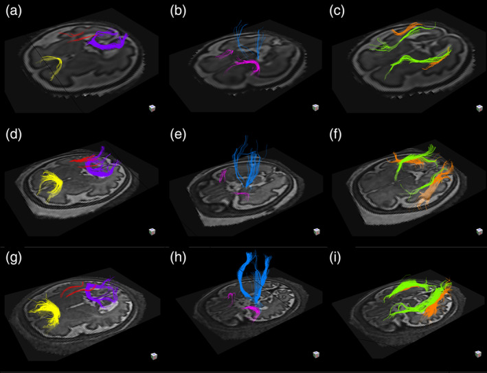
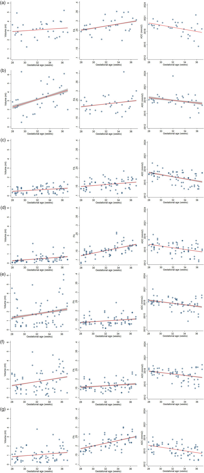
The third trimester of pregnancy is a period of rapid development of fiber bundles in the fetal white matter. Using a recently developed motion-tracked slice-to-volume registration (MT-SVR) method, we aimed to quantify tract-specific developmental changes in apparent diffusion coefficient (ADC), fractional anisotropy (FA), and volume in third trimester healthy fetuses. To this end, we reconstructed diffusion tensor images from motion corrected fetal diffusion magnetic resonance imaging data. With an approved protocol, fetal MRI exams were performed on healthy pregnant women at 3 Tesla and included multiple (2-8) diffusion scans of the fetal head (1-2 b = 0 s/mm images and 12 diffusion-sensitized images at b = 500 s/mm ). Diffusion data from 32 fetuses (13 females) with median gestational age (GA) of 33 weeks 4 days were processed with MT-SVR and deterministic tractography seeded by regions of interest corresponding to 12 major fiber tracts. Multivariable regression analysis was used to evaluate the association of GA with volume, FA, and ADC for each tract. For all tracts, the volume and FA increased, and the ADC decreased with GA. Associations reached statistical significance for: FA and ADC of the forceps major; volume and ADC for the forceps minor; FA, ADC, and volume for the cingulum; ADC, FA, and volume for the uncinate fasciculi; ADC of the inferior fronto-occipital fasciculi, ADC of the inferior longitudinal fasciculi; and FA and ADC for the corticospinal tracts. These quantitative results demonstrate the complex pattern and rates of tract-specific, GA-related microstructural changes of the developing white matter in human fetal brain.
妊娠晚期是胎儿白质中纤维束快速发育的时期。我们使用最近开发的运动跟踪切片到体素配准(MT-SVR)方法,旨在量化第三孕期健康胎儿的各向异性分数(FA)、表观扩散系数(ADC)和体积的束特异性发育变化。为此,我们从运动校正的胎儿弥散磁共振成像数据中重建了弥散张量图像。根据一项批准的方案,在 3T 对健康孕妇进行胎儿 MRI 检查,包括多次(2-8 次)胎儿头部弥散扫描(1-2 b = 0 s/mm2 的图像和 12 次 b = 500 s/mm2 的弥散敏感图像)。使用 MT-SVR 处理了 32 名胎儿(13 名女性)的弥散数据,中位数胎龄(GA)为 33 周 4 天。MT-SVR 处理和基于感兴趣区的确定性束追踪对 12 条主要纤维束进行了种子。多元回归分析用于评估 GA 与各条纤维束的体积、FA 和 ADC 的相关性。对于所有纤维束,FA 和 ADC 增加,而 ADC 随着 GA 降低。达到统计学意义的关联包括:主要钳状纤维束的 FA 和 ADC;次要钳状纤维束的体积和 ADC;胼胝体的 FA、ADC 和体积;钩束的 ADC、FA 和体积;下额枕纤维束的 ADC、下纵束的 ADC;以及皮质脊髓束的 FA 和 ADC。这些定量结果证明了人类胎儿大脑白质发育过程中束特异性、GA 相关的微观结构变化的复杂模式和速度。