Song Jae W, Gruber Gerlinde M, Patsch Janina M, Seidl Rainer, Prayer Daniela, Kasprian Gregor
Department of Radiology, Massachusetts General Hospital,, Harvard Medical School,, Boston, MA, USA.
Center for Anatomy and Cell Biology, Department of Systematic Anatomy,, Medical University of Vienna,, Vienna, Austria.
Pediatr Radiol. 2018 Apr;48(4):486-498. doi: 10.1007/s00247-017-3982-y. Epub 2018 Mar 17.
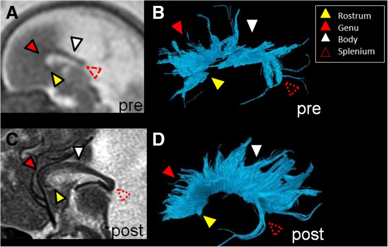
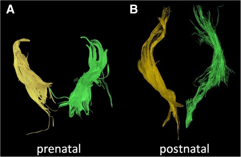
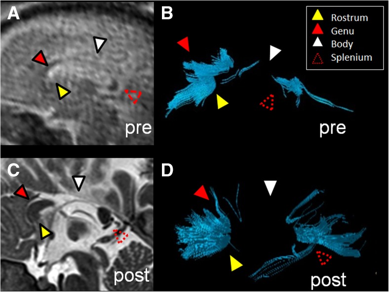
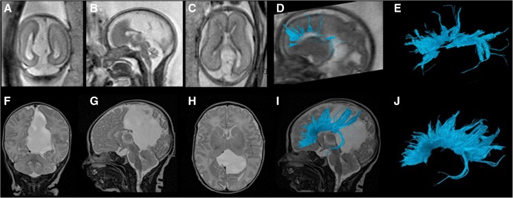
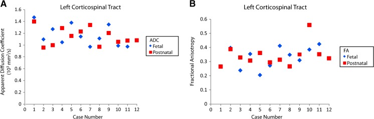
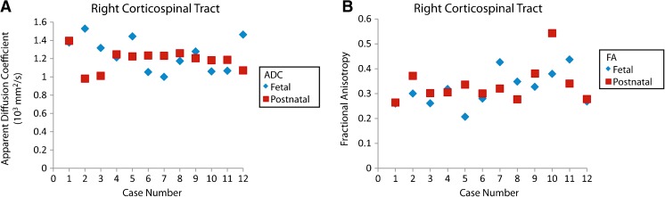
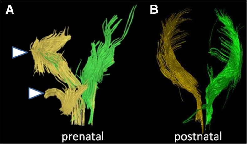
Prenatal detection of abnormal white matter tracts might serve as a structural marker for altered neurodevelopment. As a result of many technical and patient-related challenges, the accuracy of prenatal tractography remains unknown. We hypothesized that characteristics of prenatal tractography of the corpus callosum and corticospinal tracts derived from fetal diffusion tensor imaging (DTI) data are accurate and predictive of the integrity of these tracts postnatally. We compared callosal and corticospinal tracts of 12 subjects with paired prenatal (age: 23-35 gestational weeks) and postnatal (age: 1 day to 2 years) DTI examinations (b values of 0 s/mm and 700 s/mm, 16 gradient encoding directions) using deterministic tractography. Evaluation for the presence of callosal segments and corticospinal tracts showed moderate degrees of accuracy (67-75%) for the four segments of the corpus callosum and moderate to high degrees of accuracy (75-92%) for the corticospinal tracts. Positive predictive values for segments of the corpus callosum ranged from 50% to 100% and for the corticospinal tracts, 89% to 100%. Negative predictive values for segments of the corpus callosum ranged from 25% to 80% and for the corticospinal tracts, 33% to 50%. The results suggest that when the tracts are not well characterized on the fetal MR, predictions about the postnatal tracts are difficult to make. However, accounting for brain maturation, prenatal visualization of the main projection and commissural tracts can be clinically used as an important predictive tool in the context of image interpretation for the assessment of fetal brain malformations.
产前检测异常白质束可能作为神经发育改变的结构标志物。由于许多技术和患者相关的挑战,产前纤维束成像的准确性仍然未知。我们假设,从胎儿弥散张量成像(DTI)数据得出的胼胝体和皮质脊髓束的产前纤维束成像特征是准确的,并且可以预测这些束在出生后的完整性。我们使用确定性纤维束成像,比较了12名受试者配对的产前(孕周:23 - 35周)和产后(年龄:1天至2岁)DTI检查(b值为0 s/mm²和700 s/mm²,16个梯度编码方向)的胼胝体和皮质脊髓束。对胼胝体节段和皮质脊髓束的存在情况进行评估,结果显示胼胝体的四个节段准确性中等(67 - 75%),皮质脊髓束准确性为中等至高(75 - 92%)。胼胝体节段的阳性预测值范围为50%至100%,皮质脊髓束为89%至100%。胼胝体节段的阴性预测值范围为25%至80%,皮质脊髓束为33%至50%。结果表明,当胎儿磁共振成像上的束特征不明显时,很难对产后束进行预测。然而,考虑到大脑成熟情况,在评估胎儿脑畸形的图像解读中,产前对主要投射和连合束的可视化可作为一种重要的预测工具临床应用。