ZIK HALOmem, Charles-Tanford-Proteinzentrum, Martin Luther University Halle-Wittenberg, Halle/Saale, Germany.
AIMMS Division of Molecular Toxicology, Faculty of Science, Vrije Universiteit Amsterdam, Amsterdam, The Netherlands.
PLoS One. 2020 May 6;15(5):e0232540. doi: 10.1371/journal.pone.0232540. eCollection 2020.
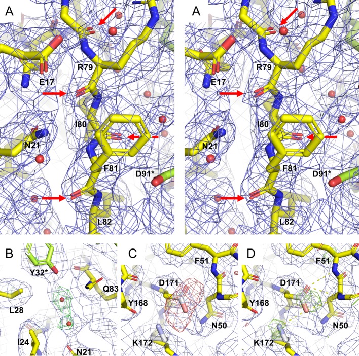
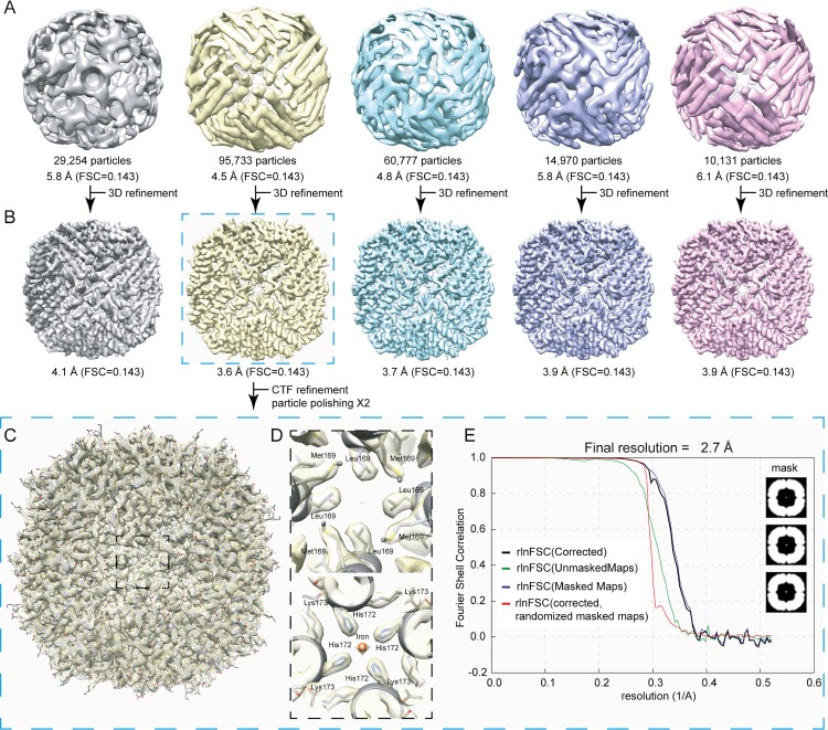
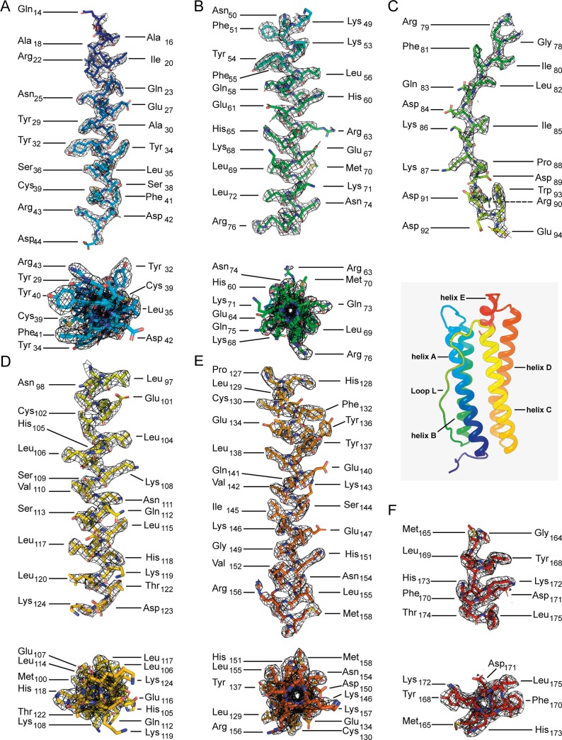
Here we present the structure of mouse H-chain apoferritin at 2.7 Å (FSC = 0.143) solved by single particle cryogenic electron microscopy (cryo-EM) using a 200 kV device, the Thermo Fisher Glacios®. This is a compact, two-lens illumination system with a constant power objective lens, without any energy filters or aberration correctors, often thought of as a "screening cryo-microscope". Coulomb potential maps reveal clear densities for main chain carbonyl oxygens, residue side chains (including alternative conformations) and bound solvent molecules. We used a quasi-crystallographic reciprocal space approach to fit model coordinates to the experimental cryo-EM map. We argue that the advantages offered by (a) the high electronic and mechanical stability of the microscope, (b) the high emission stability and low beam energy spread of the high brightness Field Emission Gun (X-FEG), (c) direct electron detection technology and (d) particle-based Contrast Transfer Function (CTF) refinement have contributed to achieving high resolution. Overall, we show that basic electron optical settings for automated cryo-electron microscopy imaging can be used to determine structures approaching atomic resolution.
在这里,我们展示了使用 200kV 设备(Thermo Fisher Glacios®)通过单颗粒低温电子显微镜(cryo-EM)解析的 2.7Å 的小鼠 H 链脱铁蛋白的结构,该分辨率下的 FSC 值为 0.143。这是一种紧凑的双透镜照明系统,配备有恒功率物镜,没有任何能量过滤器或像差校正器,通常被认为是一种“筛选 cryo-microscope”。库仑位势图显示了主链羰基氧、残基侧链(包括替代构象)和结合溶剂分子的清晰密度。我们使用准晶体倒易空间方法将模型坐标拟合到实验 cryo-EM 图中。我们认为,(a)显微镜的高电子和机械稳定性、(b)高亮度场发射枪(X-FEG)的高发射稳定性和低束能散度、(c)直接电子检测技术以及(d)基于粒子的对比度传递函数(CTF)细化的优势有助于实现高分辨率。总的来说,我们表明,自动化 cryo-EM 成像的基本电子光学设置可用于确定接近原子分辨率的结构。