• 文献检索
  • 文档翻译
  • 深度研究
  • 学术资讯
  • Suppr Zotero 插件Zotero 插件
  • 邀请有礼
  • 套餐&价格
  • 历史记录
应用&插件
Suppr Zotero 插件Zotero 插件浏览器插件Mac 客户端Windows 客户端微信小程序
定价
高级版会员购买积分包购买API积分包
服务
文献检索文档翻译深度研究API 文档MCP 服务
关于我们
关于 Suppr公司介绍联系我们用户协议隐私条款
关注我们

Suppr 超能文献

核心技术专利:CN118964589B侵权必究
粤ICP备2023148730 号-1Suppr @ 2026

文献检索

告别复杂PubMed语法,用中文像聊天一样搜索,搜遍4000万医学文献。AI智能推荐,让科研检索更轻松。

立即免费搜索

文件翻译

保留排版,准确专业,支持PDF/Word/PPT等文件格式,支持 12+语言互译。

免费翻译文档

深度研究

AI帮你快速写综述,25分钟生成高质量综述,智能提取关键信息,辅助科研写作。

立即免费体验

基于探针的共聚焦激光内镜在肺部的自动评估。

Automated evaluation of probe-based confocal laser endomicroscopy in the lung.

机构信息

Department of Radiology, University Hospital, LMU Munich, Munich, Germany.

Comprehensive Pneumology Center (CPC-M), University Hospital, LMU Munich, Helmholtz Zentrum München, Member of the German Center for Lung Research (DZL), Munich, Germany.

出版信息

PLoS One. 2020 May 6;15(5):e0232847. doi: 10.1371/journal.pone.0232847. eCollection 2020.

DOI:10.1371/journal.pone.0232847
PMID:32374768
原文链接:https://pmc.ncbi.nlm.nih.gov/articles/PMC7202624/
Abstract

RATIONALE

Probe-based confocal endomicroscopy provides real time videos of autoflourescent elastin structures within the alveoli. With it, multiple changes in the elastin structure due to different diffuse parenchymal lung diseases have previously been described. However, these evaluations have mainly relied on qualitative evaluation by the examiner and manually selected parts post-examination.

OBJECTIVES

To develop a fully automatic method for quantifying structural properties of the imaged alveoli elastin and to perform a preliminary assessment of their diagnostic potential.

METHODS

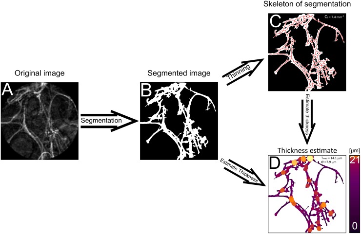
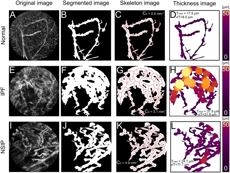
46 patients underwent probe-based confocal endomicroscopy, of which 38 were divided into 4 groups categorizing different diffuse parenchymal lung diseases. 8 patients were imaged in representative healthy lung areas and used as control group. Alveolar elastin structures were automatically segmented with a trained machine learning algorithm and subsequently evaluated with two methods developed for quantifying the local thickness and structural connectivity.

MEASUREMENTS AND MAIN RESULTS

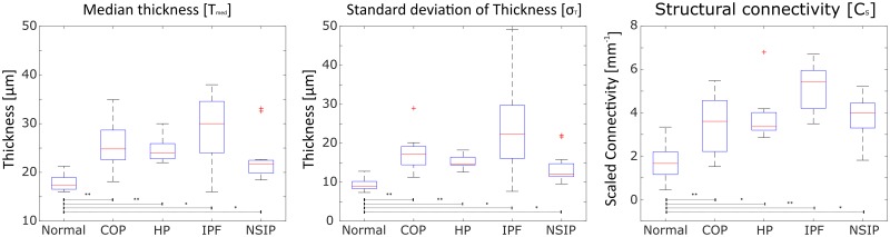
The automatic segmentation algorithm performed generally well and all 4 patient groups showed statistically significant differences with median elastin thickness, standard deviation of thickness and connectivity compared to the control group.

CONCLUSION

Alveoli elastin structures can be quantified based on their structural connectivity and thickness statistics with a fully-automated algorithm and initial results highlight its potential for distinguishing parenchymal lung diseases from normal alveoli.

摘要

原理

基于探针的共聚焦内窥镜提供了肺泡内自发荧光弹性结构的实时视频。利用它,先前已经描述了由于不同弥漫性实质性肺疾病导致的弹性结构的多种变化。然而,这些评估主要依赖于检查者的定性评估以及检查后的手动选择部分。

目的

开发一种定量分析成像肺泡弹性蛋白结构特性的全自动方法,并对其诊断潜力进行初步评估。

方法

46 名患者接受了基于探针的共聚焦内窥镜检查,其中 38 名患者分为 4 组,分类为不同的弥漫性实质性肺疾病。8 名患者在代表性的健康肺区进行成像,作为对照组。使用经过训练的机器学习算法对肺泡弹性结构进行自动分割,然后使用两种方法对局部厚度和结构连通性进行评估。

测量和主要结果

自动分割算法总体表现良好,与对照组相比,所有 4 组患者的弹性蛋白厚度中位数、厚度标准差和连通性均有统计学显著差异。

结论

可以使用全自动算法基于结构连通性和厚度统计数据对肺泡弹性结构进行定量分析,初步结果突出了其区分实质肺疾病与正常肺泡的潜力。

https://cdn.ncbi.nlm.nih.gov/pmc/blobs/37d7/7202624/dbcafea71035/pone.0232847.g003.jpg
https://cdn.ncbi.nlm.nih.gov/pmc/blobs/37d7/7202624/be3e573b2d85/pone.0232847.g001.jpg
https://cdn.ncbi.nlm.nih.gov/pmc/blobs/37d7/7202624/bf4ad25a5b54/pone.0232847.g002.jpg
https://cdn.ncbi.nlm.nih.gov/pmc/blobs/37d7/7202624/dbcafea71035/pone.0232847.g003.jpg
https://cdn.ncbi.nlm.nih.gov/pmc/blobs/37d7/7202624/be3e573b2d85/pone.0232847.g001.jpg
https://cdn.ncbi.nlm.nih.gov/pmc/blobs/37d7/7202624/bf4ad25a5b54/pone.0232847.g002.jpg
https://cdn.ncbi.nlm.nih.gov/pmc/blobs/37d7/7202624/dbcafea71035/pone.0232847.g003.jpg

相似文献

1
Automated evaluation of probe-based confocal laser endomicroscopy in the lung.基于探针的共聚焦激光内镜在肺部的自动评估。
PLoS One. 2020 May 6;15(5):e0232847. doi: 10.1371/journal.pone.0232847. eCollection 2020.
2
Probe-based confocal laser endomicroscopy in diagnosis of desquamative interstitial pneumonia in nonsmoker.基于探头的共聚焦激光内镜检查在非吸烟者脱屑性间质性肺炎诊断中的应用
Monaldi Arch Chest Dis. 2019 Sep 10;89(3). doi: 10.4081/monaldi.2019.1052.
3
Fluorescein-aided confocal laser endomicroscopy of the lung.肺的荧光素辅助共聚焦激光内镜检查。
Respiration. 2011;81(1):32-8. doi: 10.1159/000320365. Epub 2010 Aug 21.
4
Confocal Laser Endomicroscopy as a Guidance Tool for Transbronchial Lung Cryobiopsies in Interstitial Lung Disorder.共聚焦激光支气管镜肺活检术在间质性肺疾病中的应用
Respiration. 2019;97(3):259-263. doi: 10.1159/000493271. Epub 2018 Nov 14.
5
Probe-based confocal laser endomicroscopy of the respiratory tract: a data consistency analysis.探头式共聚焦激光支气管镜检查:数据一致性分析。
Respir Med. 2013 Aug;107(8):1234-40. doi: 10.1016/j.rmed.2013.04.018. Epub 2013 May 23.
6
Confocal laser endomicroscopy for diagnosing lung cancer in vivo.共聚焦激光内镜检查术用于体内诊断肺癌。
Eur Respir J. 2013 Jun;41(6):1401-8. doi: 10.1183/09031936.00062512. Epub 2012 Sep 20.
7
In vivo probe-based confocal laser endomicroscopy in chronic interstitial lung diseases: Specific descriptors and correlation with chest CT.体内探针式共聚焦激光内镜检查在慢性间质性肺疾病中的应用:特定描述符与胸部 CT 的相关性。
Respirology. 2019 Aug;24(8):783-791. doi: 10.1111/resp.13507. Epub 2019 Feb 27.
8
Probe-based confocal laser endomicroscopy in COVID-19.基于探头的共聚焦激光内镜检查在 COVID-19 中的应用。
Adv Respir Med. 2021;89(4):456-459. doi: 10.5603/ARM.a2021.0067. Epub 2021 Jul 16.
9
The microanatomy of the alveolar duct of the human lung imaged by confocal microscopy and visualised with computer-based 3D reconstruction.
Comput Med Imaging Graph. 1993 May-Jun;17(3):201-10. doi: 10.1016/0895-6111(93)90044-n.
10
Comparison of in vivo probe-based confocal laser endomicroscopy with histopathology in lung cancer: A move toward optical biopsy.肺癌中基于探头的共聚焦激光内镜检查与组织病理学的体内比较:迈向光学活检的一步。
Respirology. 2015 Aug;20(6):967-74. doi: 10.1111/resp.12578. Epub 2015 Jun 19.

引用本文的文献

1
Ex vivo use of probe-based confocal laser endomicroscopy in lung cancer visualization: an ex vivo pilot study.基于探头的共聚焦激光内镜术在肺癌可视化中的体外应用:一项体外初步研究。
Rom J Morphol Embryol. 2025 Jan-Mar;66(1):163-171. doi: 10.47162/RJME.66.1.15.
2
The cryobiopsy in interstitial lung diseases guided by probe-based confocal laser endomicroscopy is feasible.探针式共聚焦激光显微内镜引导下的肺间质疾病冷冻活检是可行的。
Clin Respir J. 2023 Oct;17(10):998-1005. doi: 10.1111/crj.13669. Epub 2023 Aug 16.
3
The role of confocal laser endomicroscopy in pulmonary medicine.

本文引用的文献

1
In vivo probe-based confocal laser endomicroscopy in chronic interstitial lung diseases: Specific descriptors and correlation with chest CT.体内探针式共聚焦激光内镜检查在慢性间质性肺疾病中的应用:特定描述符与胸部 CT 的相关性。
Respirology. 2019 Aug;24(8):783-791. doi: 10.1111/resp.13507. Epub 2019 Feb 27.
2
Confocal Laser Endomicroscopy as a Guidance Tool for Transbronchial Lung Cryobiopsies in Interstitial Lung Disorder.共聚焦激光支气管镜肺活检术在间质性肺疾病中的应用
Respiration. 2019;97(3):259-263. doi: 10.1159/000493271. Epub 2018 Nov 14.
3
Taking Another View on Lung Fibrosis.
共聚焦激光内镜在肺部医学中的应用。
Eur Respir Rev. 2023 Jan 25;32(167). doi: 10.1183/16000617.0185-2022. Print 2023 Mar 31.
4
Bronchoscopic Probe-Based Confocal Laser Endomicroscopy to Diagnose Diffuse Parenchymal Lung Diseases.基于支气管镜探头的共聚焦激光内镜检查用于诊断弥漫性肺实质疾病。
Sarcoidosis Vasc Diffuse Lung Dis. 2022;39(2):e2022016. doi: 10.36141/svdld.v39i2.11280. Epub 2022 Jun 29.
5
Diagnosis of visceral pleural invasion using confocal laser endomicroscopy during lung cancer surgery.肺癌手术中使用共聚焦激光内镜显微镜诊断脏层胸膜侵犯
J Thorac Dis. 2021 Aug;13(8):4742-4752. doi: 10.21037/jtd-21-137.
6
Solitary pulmonary nodule imaging approaches and the role of optical fibre-based technologies.孤立性肺结节的成像方法及基于光纤技术的作用。
Eur Respir J. 2021 Mar 25;57(3). doi: 10.1183/13993003.02537-2020. Print 2021 Mar.
对肺纤维化的另一种看法。
Am J Respir Crit Care Med. 2018 Apr 1;197(7):947-948. doi: 10.1164/rccm.201708-1683IM.
4
Transbronchial Cryobiopsies for the Diagnosis of Diffuse Parenchymal Lung Diseases: Expert Statement from the Cryobiopsy Working Group on Safety and Utility and a Call for Standardization of the Procedure.经支气管冷冻活检诊断弥漫性实质性肺疾病:安全与效用冷冻活检工作组专家声明及对该操作标准化的呼吁
Respiration. 2018;95(3):188-200. doi: 10.1159/000484055. Epub 2018 Jan 9.
5
Diagnostic criteria for idiopathic pulmonary fibrosis: a Fleischner Society White Paper.特发性肺纤维化的诊断标准:弗利彻协会白皮书。
Lancet Respir Med. 2018 Feb;6(2):138-153. doi: 10.1016/S2213-2600(17)30433-2. Epub 2017 Nov 15.
6
Trainable Weka Segmentation: a machine learning tool for microscopy pixel classification.可训练的 WEKA 分割:一种用于显微镜像素分类的机器学习工具。
Bioinformatics. 2017 Aug 1;33(15):2424-2426. doi: 10.1093/bioinformatics/btx180.
7
Fibred confocal fluorescence microscopy in the diagnosis of interstitial lung diseases.纤维共聚焦荧光显微镜在间质性肺疾病诊断中的应用
J Thorac Dis. 2016 Dec;8(12):3505-3514. doi: 10.21037/jtd.2016.12.60.
8
The diagnosis of idiopathic pulmonary fibrosis: current and future approaches.特发性肺纤维化的诊断:当前和未来的方法。
Lancet Respir Med. 2017 Jan;5(1):61-71. doi: 10.1016/S2213-2600(16)30325-3. Epub 2016 Dec 6.
9
Acute lung allograft rejection: diagnostic role of probe-based confocal laser endomicroscopy of the respiratory tract.急性肺移植排斥反应:呼吸道探针式共聚焦激光内镜检查的诊断作用。
J Heart Lung Transplant. 2014 May;33(5):492-8. doi: 10.1016/j.healun.2014.01.857. Epub 2014 Jan 24.
10
Probe-based confocal laser endomicroscopy of the respiratory tract: a data consistency analysis.探头式共聚焦激光支气管镜检查:数据一致性分析。
Respir Med. 2013 Aug;107(8):1234-40. doi: 10.1016/j.rmed.2013.04.018. Epub 2013 May 23.