Translational Genomics Research Institute, Phoenix, AZ, USA.
Surgery/Ophthalmology, USA; NMVA Health Care System, Albuquerque, NM, USA.
Exp Eye Res. 2020 Jun;195:108043. doi: 10.1016/j.exer.2020.108043. Epub 2020 May 4.
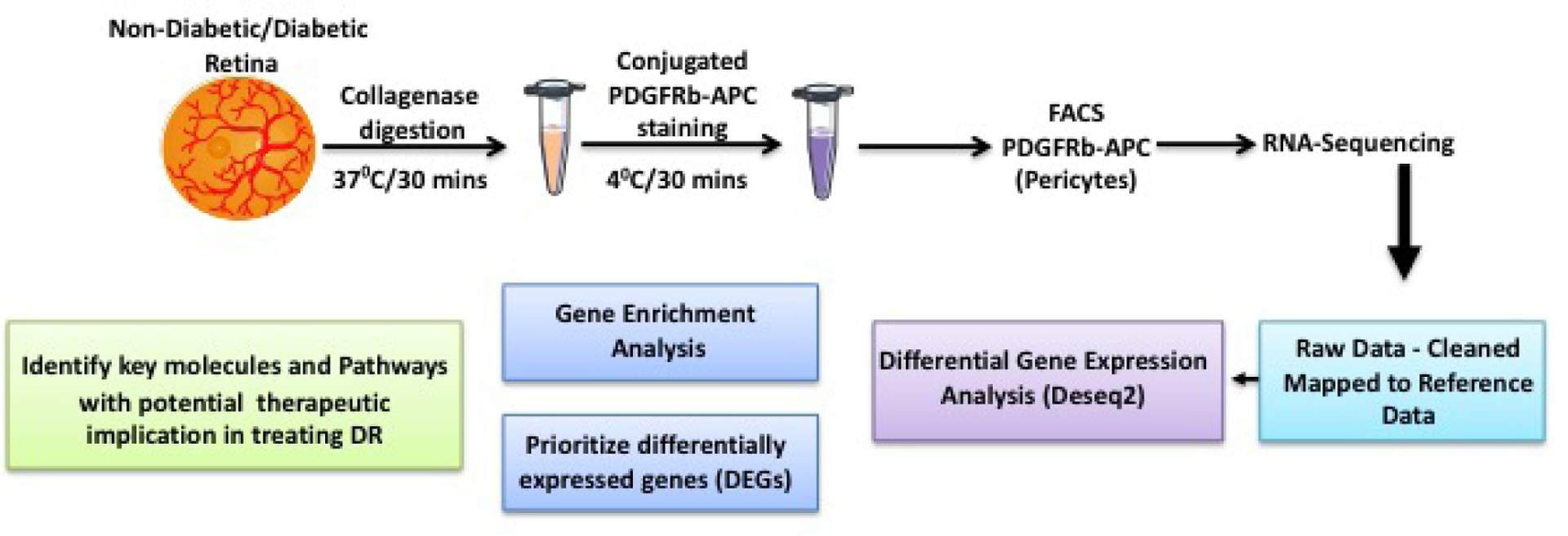
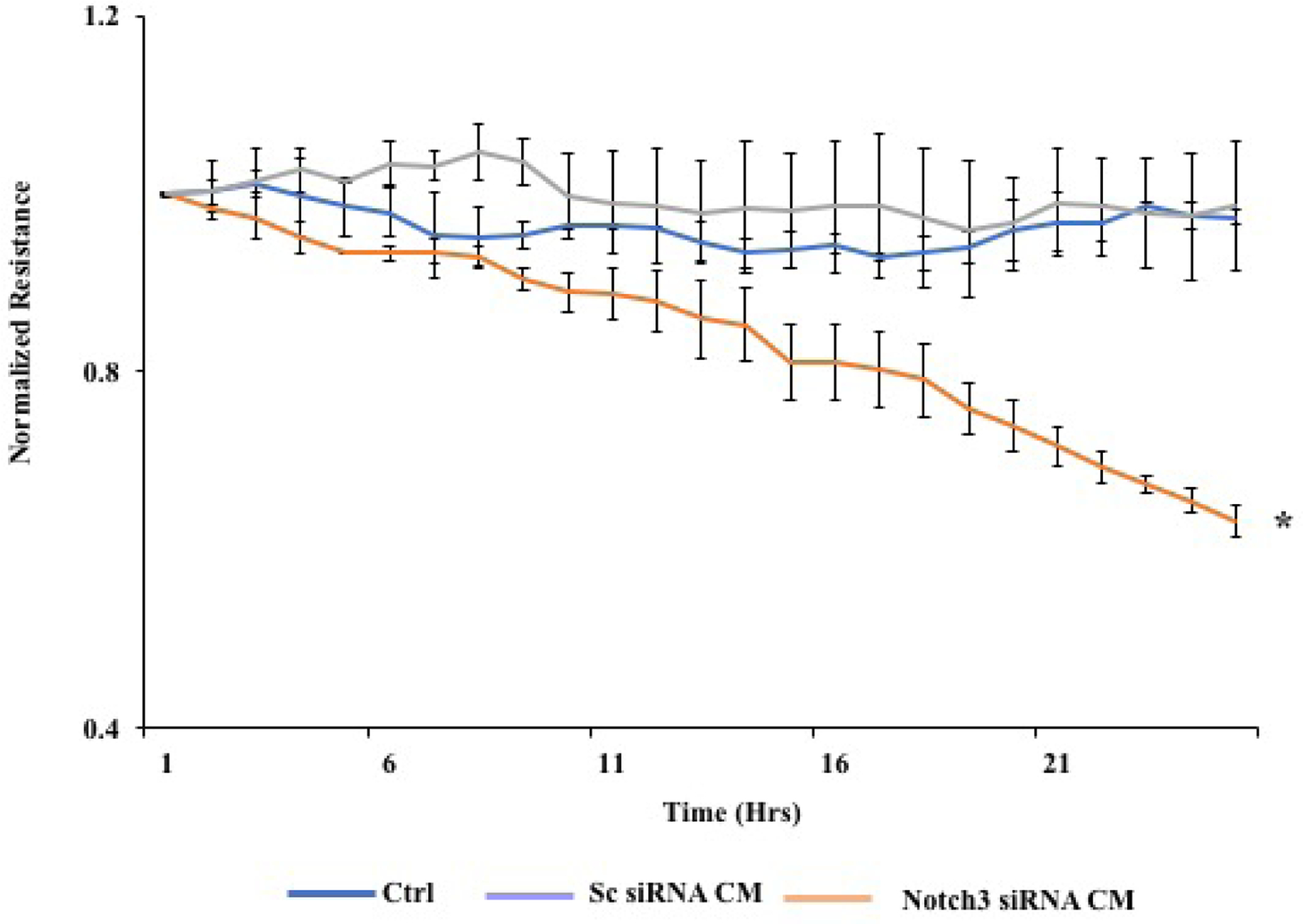
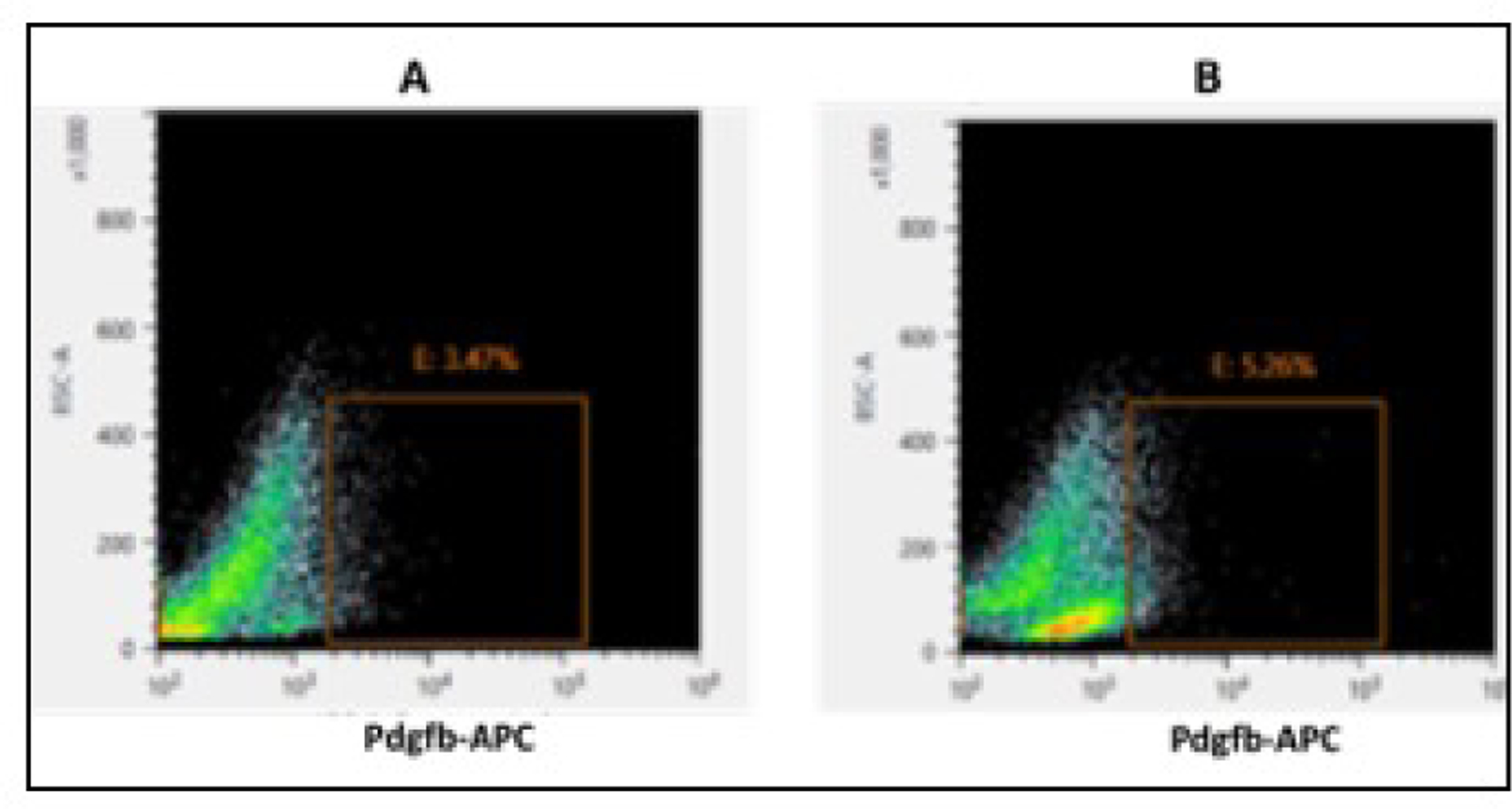
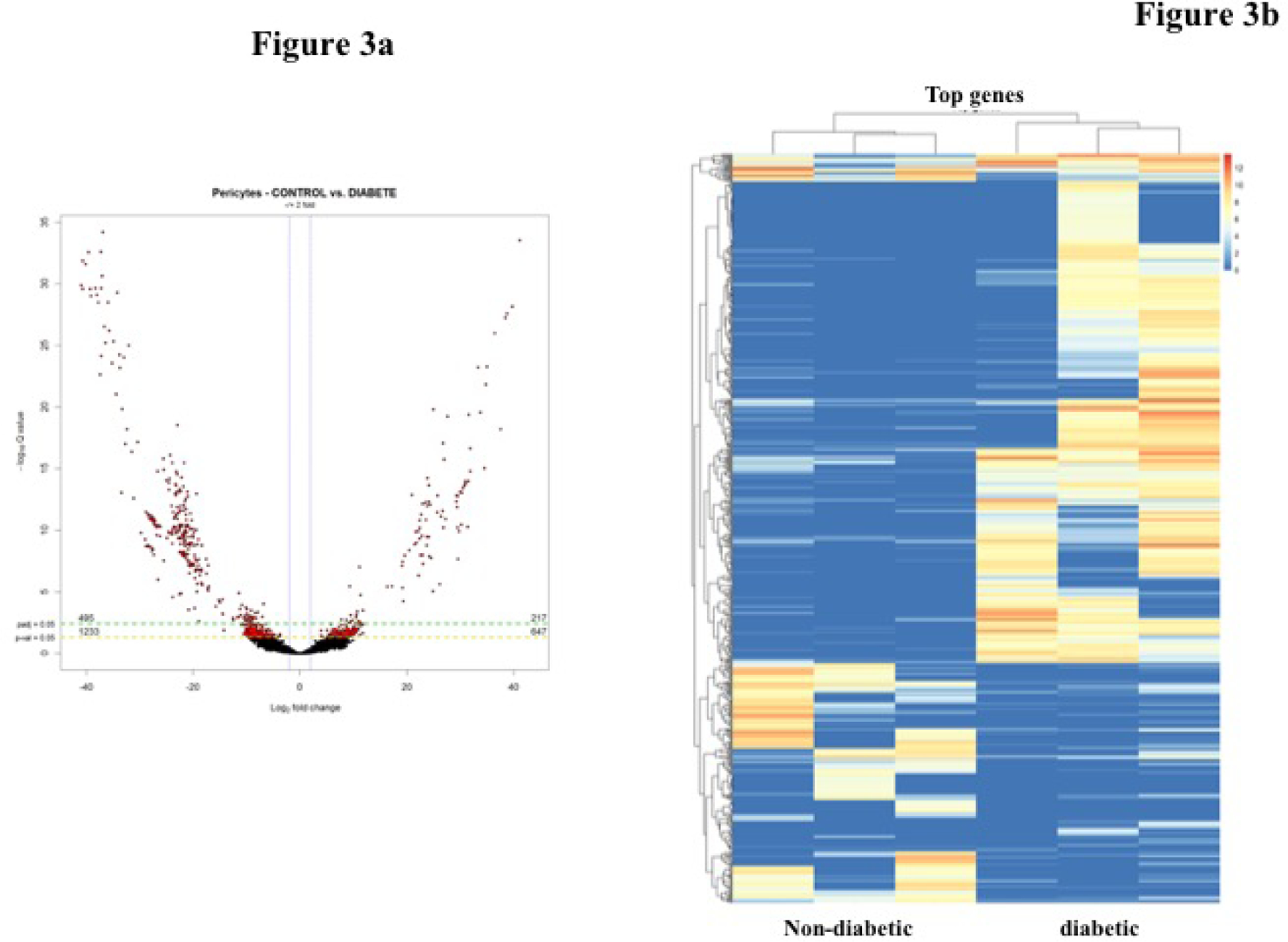
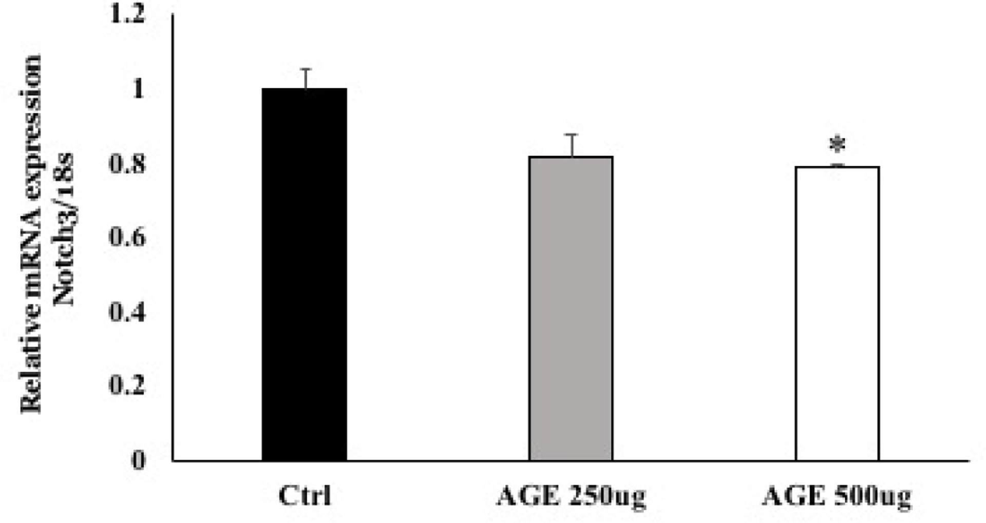
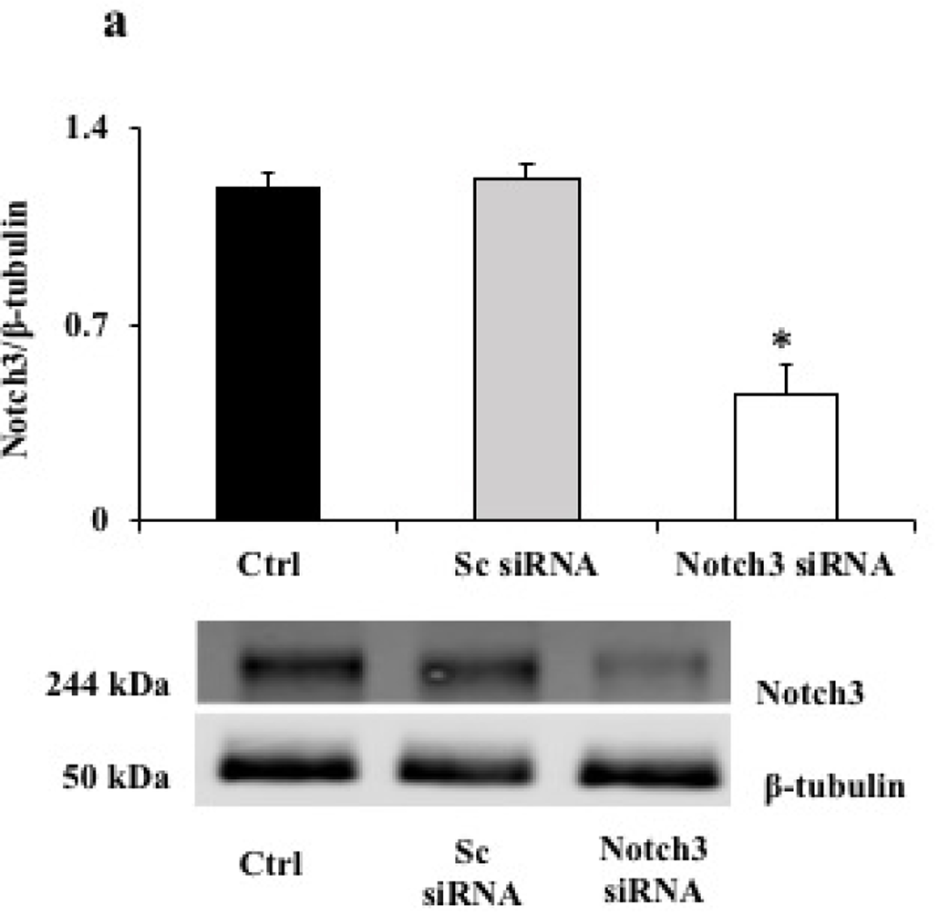
Selective pericyte loss, the histological hallmark of early diabetic retinopathy (DR), enhances the breakdown of the blood-retinal barrier (BRB) in diabetes. However, the role of pericytes on BRB alteration in diabetes and the signaling pathways involved in their effects are currently unknown. To understand the role of diabetes-induced molecular alteration of pericytes, we performed transcriptomic analysis of sorted retinal pericytes from mice model of diabetes. Retinal tissue from non-diabetic and diabetic (duration 3 months) mouse eyes (n = 10 in each group) were used to isolate pericytes through fluorescent activated cell sorting (FACS) using pericyte specific fluorescent antibodies, PDGFRb-APC. For RNA sequencing and qPCR analysis, a cDNA library was generated using template switching oligo and the resulting libraries were sequenced using paired-end Illumina sequencing. Molecular functional pathways were analyzed using differentially expressed genes (DEGs). Differential expression analysis revealed 217 genes significantly upregulated and 495 genes downregulated, in pericytes isolated from diabetic animals. These analyses revealed a core set of differentially expressed genes that could potentially contribute to the pericyte dysfunction in diabetes and highlighted the pattern of functional connectivity between key candidate genes and blood retinal barrier alteration mechanisms. The top up-regulated gene list included: Ext2, B3gat3, Gpc6, Pip5k1c and Pten and down-regulated genes included: Notch3, Xbp1, Gpc4, Atp1a2 and AKT3. Out of these genes, we further validated one of the down regulated genes, Notch 3 and its role in BRB alteration in diabetic retinopathy. We confirmed the downregulation of Notch3 expression in human retinal pericytes exposed to Advanced Glycation End-products (AGEs) treatment mimicking the chronic hyperglycemia effect. Exploration of pericyte-conditioned media demonstrated that loss of NOTCH3 in pericyte led to increased permeability of endothelial cell monolayers. Collectively, we identify a role for NOTCH3 in pericyte dysfunction in diabetes. Further validation of other DEGs to identify cell specific molecular change through whole transcriptomic approach in diabetic retina will provide novel insight into the pathogenesis of DR and novel therapeutic targets.
选择性周细胞丢失是早期糖尿病视网膜病变(DR)的组织学标志,可增强糖尿病中血视网膜屏障(BRB)的破坏。然而,周细胞在糖尿病中 BRB 改变中的作用及其涉及的信号通路目前尚不清楚。为了了解糖尿病诱导的周细胞分子改变的作用,我们对糖尿病小鼠模型中分离的视网膜周细胞进行了转录组分析。使用荧光激活细胞分选(FACS),通过周细胞特异性荧光抗体 PDGFRb-APC 从非糖尿病和糖尿病(持续 3 个月)小鼠眼睛的视网膜组织中(每组 10 只)分离周细胞。为了进行 RNA 测序和 qPCR 分析,使用模板转换寡核苷酸生成 cDNA 文库,然后使用配对末端 Illumina 测序对文库进行测序。使用差异表达基因(DEGs)分析分子功能途径。差异表达分析显示,在从糖尿病动物中分离的周细胞中,有 217 个基因显著上调,495 个基因下调。这些分析揭示了一组潜在的差异表达基因,这些基因可能有助于糖尿病中的周细胞功能障碍,并突出了关键候选基因与血视网膜屏障改变机制之间的功能连接模式。上调基因列表的前 10 个基因包括:Ext2、B3gat3、Gpc6、Pip5k1c 和 Pten,下调基因包括:Notch3、Xbp1、Gpc4、Atp1a2 和 AKT3。在这些基因中,我们进一步验证了下调基因之一 Notch3 在糖尿病性视网膜病变中对 BRB 改变的作用。我们证实了在暴露于模拟慢性高血糖作用的晚期糖基化终产物(AGEs)的人视网膜周细胞中 Notch3 表达下调。对周细胞条件培养基的探索表明,周细胞中 NOTCH3 的丢失导致内皮细胞单层的通透性增加。总之,我们确定了 NOTCH3 在糖尿病中周细胞功能障碍中的作用。通过全转录组方法在糖尿病视网膜中进一步验证其他 DEGs,以确定细胞特异性分子变化,将为 DR 的发病机制和新的治疗靶点提供新的见解。