Oba Sibel, Işıl Canan Tülay, Türk Hacer Şebnem, Karamürsel Sacit, Aksu Serkan, Kaba Meltem, Kılınç Leyla, Dokucu Ali Ihsan
Department of Anestesiology and Reanimation, Sisli Hamidiye Etfal Training and Research Hospital, Istanbul, Turkey.
Department of Physiology, Istinye University Faculty of Medicine, Istanbul, Turkey.
Sisli Etfal Hastan Tip Bul. 2019 Aug 26;53(3):284-289. doi: 10.14744/SEMB.2018.59454. eCollection 2019.
Anesthetic applications may cause increased neuronal damage in infants and children. Commonly cognitive or learning disability tests were used to investigate the neurological progress in children. Visual Evoked Potential is a gross electrical signal generated by the occipital regions of the cerebral cortex in response to visual stimulation and an objective assessment of brain function. In this study, to acquire more objective results, Visual Evoked Potential responses of children who had multiple exposures to anesthesia during the treatment of corrosive esophagitis were compared to children who have never received anesthesia before.
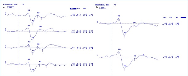
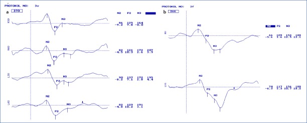
In this prospective, single-blinded, randomized, controlled study, 25 children, who were admitted to our pediatric surgery clinic because of corrosive esophagitis and who received general anesthesia more than 15 times composed Group-P; 25 children, who admitted to our well-child-clinic and who had never received anesthesia before consisted Group-C. The flash and pattern VEP responses of both groups were measured at the electrophysiology laboratory without any anesthetic drug application. The VEP responses of children in Group-P were recorded at least three days after the last exposure to anesthesia.
Latencies and amplitudes of the N2 and P2 components of the pattern and flash VEP responses were statistically significantly different between the two groups (p=0.000).
This study shows that in children who had repeated anesthetic applications VEP parameters are significantly altered. We believe that VEP responses may be a reliable objective criterion for the evaluation of anesthesia neurotoxicity.
麻醉应用可能会增加婴幼儿和儿童的神经元损伤。通常使用认知或学习能力测试来研究儿童的神经发育进程。视觉诱发电位是大脑皮层枕叶区域对视觉刺激产生的一种总体电信号,是对脑功能的一种客观评估。在本研究中,为了获得更客观的结果,将在腐蚀性食管炎治疗期间多次接受麻醉的儿童的视觉诱发电位反应与从未接受过麻醉的儿童进行了比较。
在这项前瞻性、单盲、随机对照研究中,因腐蚀性食管炎入住我们儿科外科门诊且接受全身麻醉超过15次的25名儿童组成P组;入住我们儿童健康门诊且从未接受过麻醉的25名儿童组成C组。在电生理实验室,在未应用任何麻醉药物的情况下测量两组儿童的闪光视觉诱发电位和图形视觉诱发电位反应。P组儿童的视觉诱发电位反应在最后一次接触麻醉至少三天后记录。
两组间图形视觉诱发电位和闪光视觉诱发电位反应的N2和P2成分的潜伏期和波幅在统计学上有显著差异(p = 0.000)。
本研究表明,在反复接受麻醉的儿童中,视觉诱发电位参数有显著改变。我们认为,视觉诱发电位反应可能是评估麻醉神经毒性的一个可靠客观标准。