Hayashi Hironobu, Kawaguchi Masahiko
Department of Anesthesiology, Nara Medical University, Kashihara, Nara, Japan.
Korean J Anesthesiol. 2017 Apr;70(2):127-135. doi: 10.4097/kjae.2017.70.2.127. Epub 2017 Mar 6.
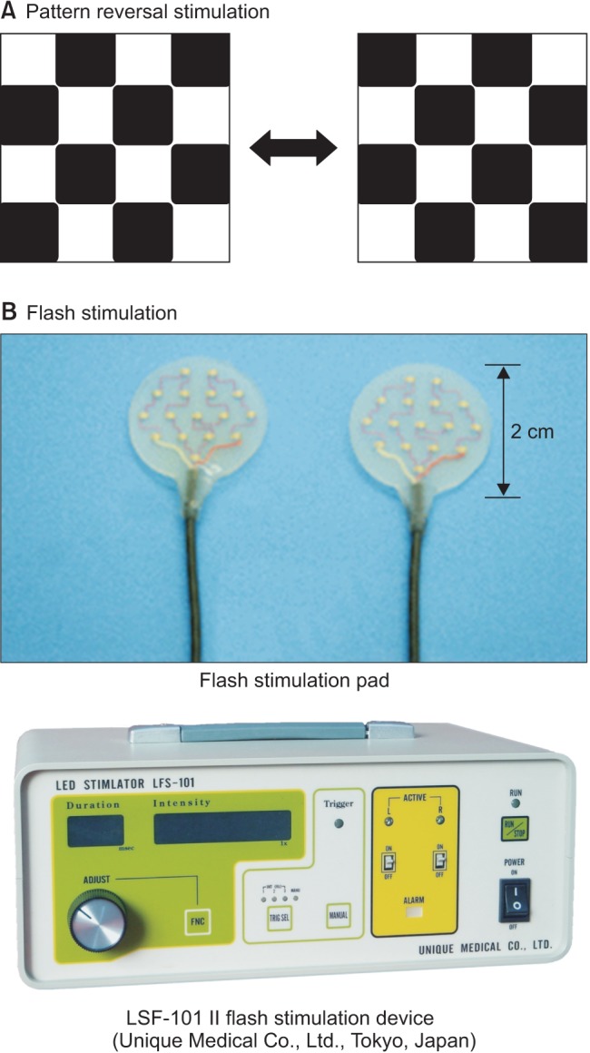
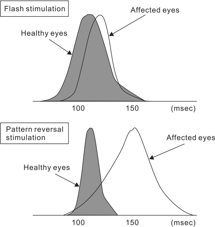
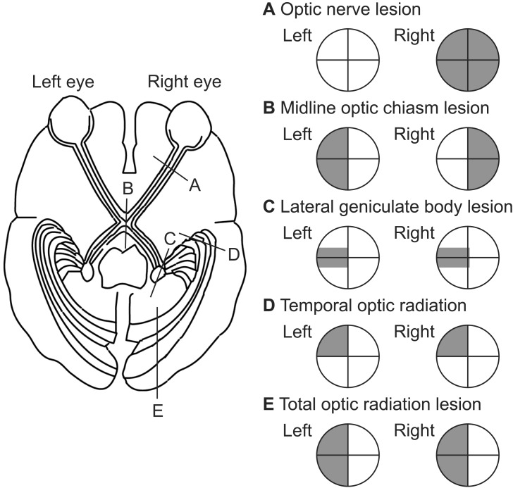
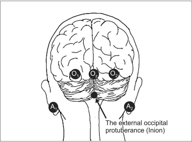
In neurosurgical procedures that may cause visual impairment in the intraoperative period, the monitoring of flash visual evoked potential (VEP) is clinically used to evaluate visual function. Patients are unconscious during surgery under general anesthesia, making flash VEP monitoring useful as it can objectively evaluate visual function. The flash stimulus input to the retina is transmitted to the optic nerve, optic chiasm, optic tract, lateral geniculate body, optic radiation (geniculocalcarine tract), and visual cortical area, and the VEP waveform is recorded from the occipital region. Intraoperative flash VEP monitoring allows detection of dysfunction arising anywhere in the optic pathway, from the retina to the visual cortex. Particularly important steps to obtain reproducible intraoperative flash VEP waveforms under general anesthesia are total intravenous anesthesia with propofol, use of retinal flash stimulation devices using high-intensity light-emitting diodes, and a combination of electroretinography to confirm that the flash stimulus has reached the retina. Relatively major postoperative visual impairment can be detected by intraoperative decreases in the flash VEP amplitude.
在可能导致术中视力损害的神经外科手术中,闪光视觉诱发电位(VEP)监测在临床上用于评估视觉功能。患者在全身麻醉下手术时处于无意识状态,这使得闪光VEP监测很有用,因为它可以客观地评估视觉功能。输入视网膜的闪光刺激会传输至视神经、视交叉、视束、外侧膝状体、视辐射(膝距束)和视觉皮层区域,并从枕部记录VEP波形。术中闪光VEP监测能够检测从视网膜到视觉皮层的视神经通路中任何部位出现的功能障碍。在全身麻醉下获得可重复的术中闪光VEP波形的特别重要步骤包括使用丙泊酚进行全静脉麻醉、使用采用高强度发光二极管的视网膜闪光刺激装置以及结合视网膜电图来确认闪光刺激已到达视网膜。术中闪光VEP振幅降低可检测出相对严重的术后视力损害。