• 文献检索
  • 文档翻译
  • 深度研究
  • 学术资讯
  • Suppr Zotero 插件Zotero 插件
  • 邀请有礼
  • 套餐&价格
  • 历史记录
应用&插件
Suppr Zotero 插件Zotero 插件浏览器插件Mac 客户端Windows 客户端微信小程序
定价
高级版会员购买积分包购买API积分包
服务
文献检索文档翻译深度研究API 文档MCP 服务
关于我们
关于 Suppr公司介绍联系我们用户协议隐私条款
关注我们

Suppr 超能文献

核心技术专利:CN118964589B侵权必究
粤ICP备2023148730 号-1Suppr @ 2026

文献检索

告别复杂PubMed语法,用中文像聊天一样搜索,搜遍4000万医学文献。AI智能推荐,让科研检索更轻松。

立即免费搜索

文件翻译

保留排版,准确专业,支持PDF/Word/PPT等文件格式,支持 12+语言互译。

免费翻译文档

深度研究

AI帮你快速写综述,25分钟生成高质量综述,智能提取关键信息,辅助科研写作。

立即免费体验

Ena/VASP 的缺失会干扰片状伪足的结构、运动和整合素依赖性黏附。

Loss of Ena/VASP interferes with lamellipodium architecture, motility and integrin-dependent adhesion.

机构信息

Institute for Biophysical Chemistry, Hannover Medical School, Hannover, Germany.

CytoMorphoLab, Laboratoire de Physiologie cellulaire et Végétale, Interdisciplinary ResearchInstitute of Grenoble, CEA, CNRS, INRA, Grenoble-Alpes University, Grenoble, France.

出版信息

Elife. 2020 May 11;9:e55351. doi: 10.7554/eLife.55351.

DOI:10.7554/eLife.55351
PMID:32391788
原文链接:https://pmc.ncbi.nlm.nih.gov/articles/PMC7239657/
Abstract

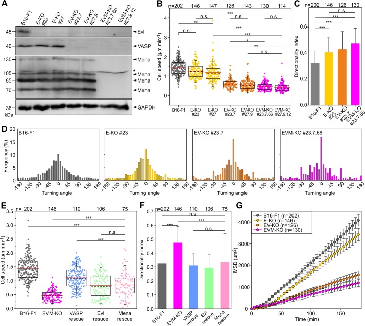
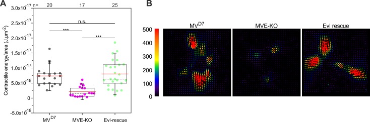
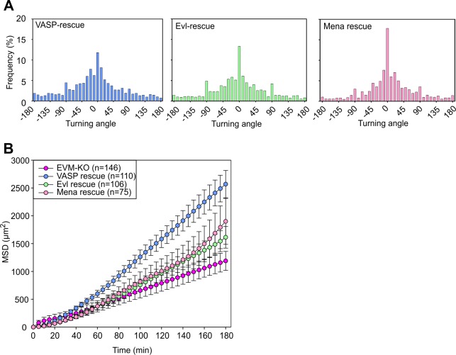
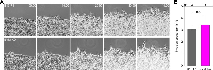
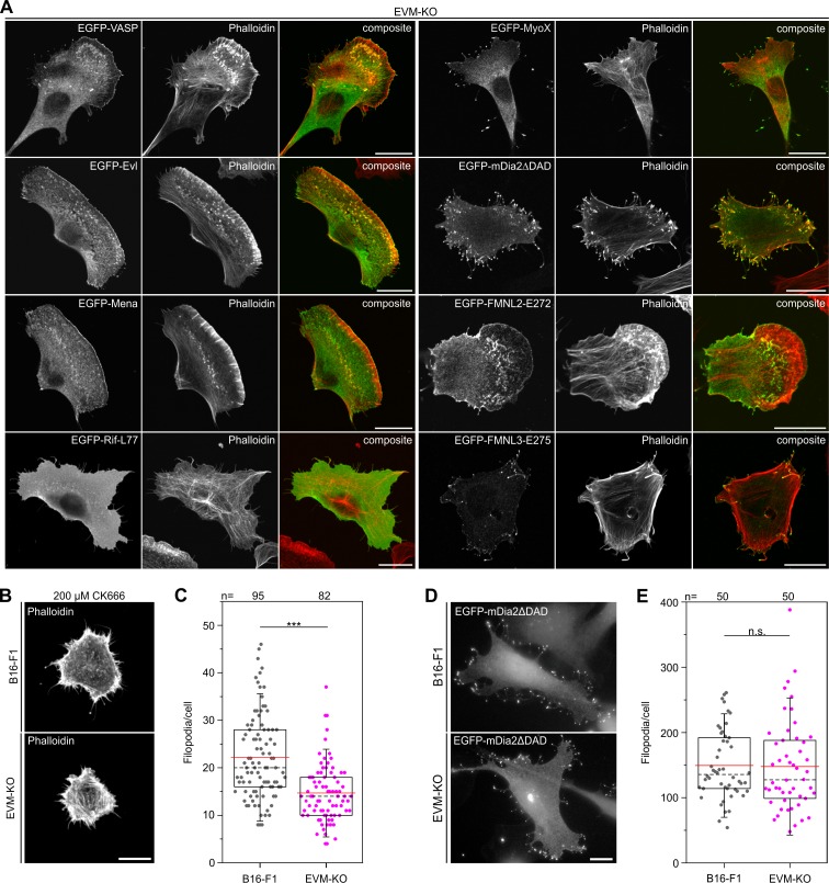
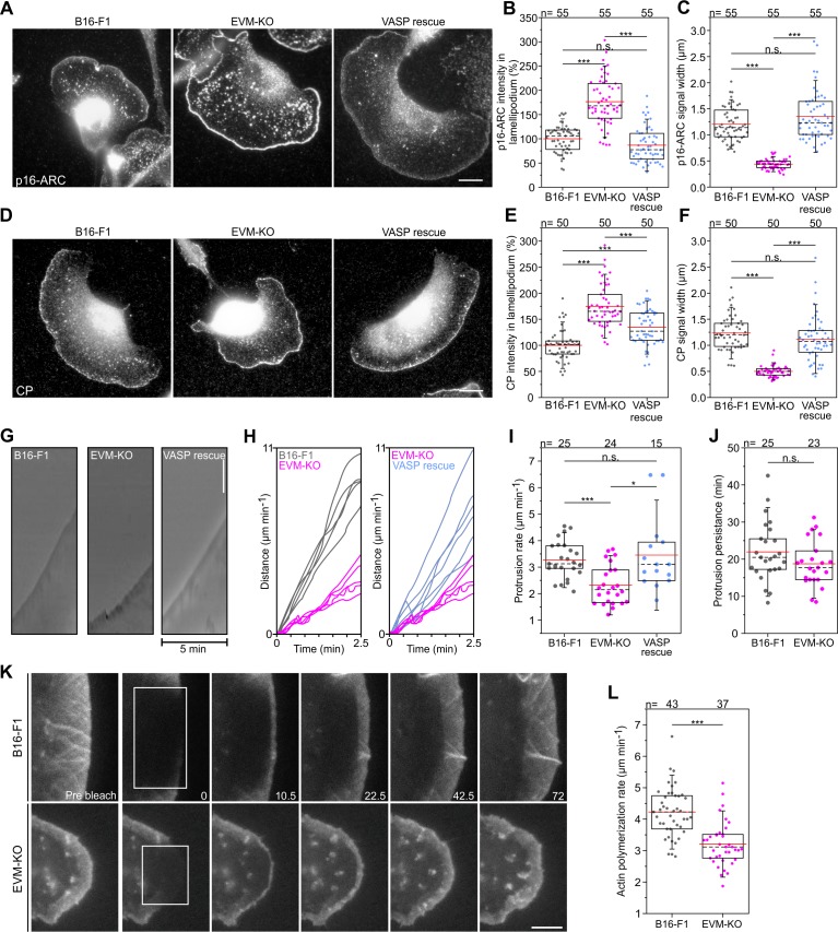
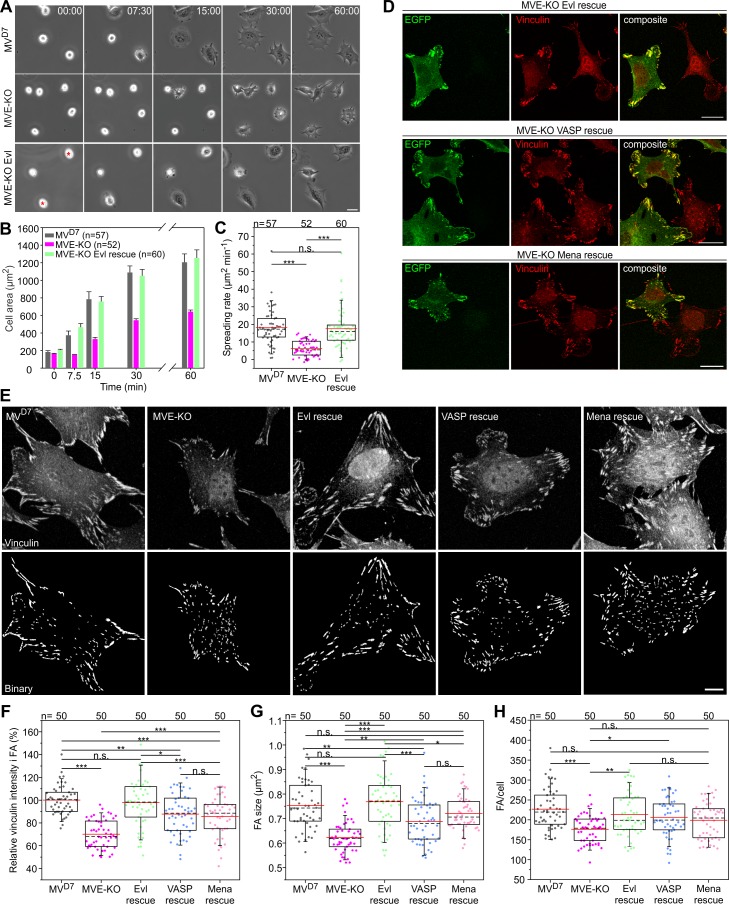
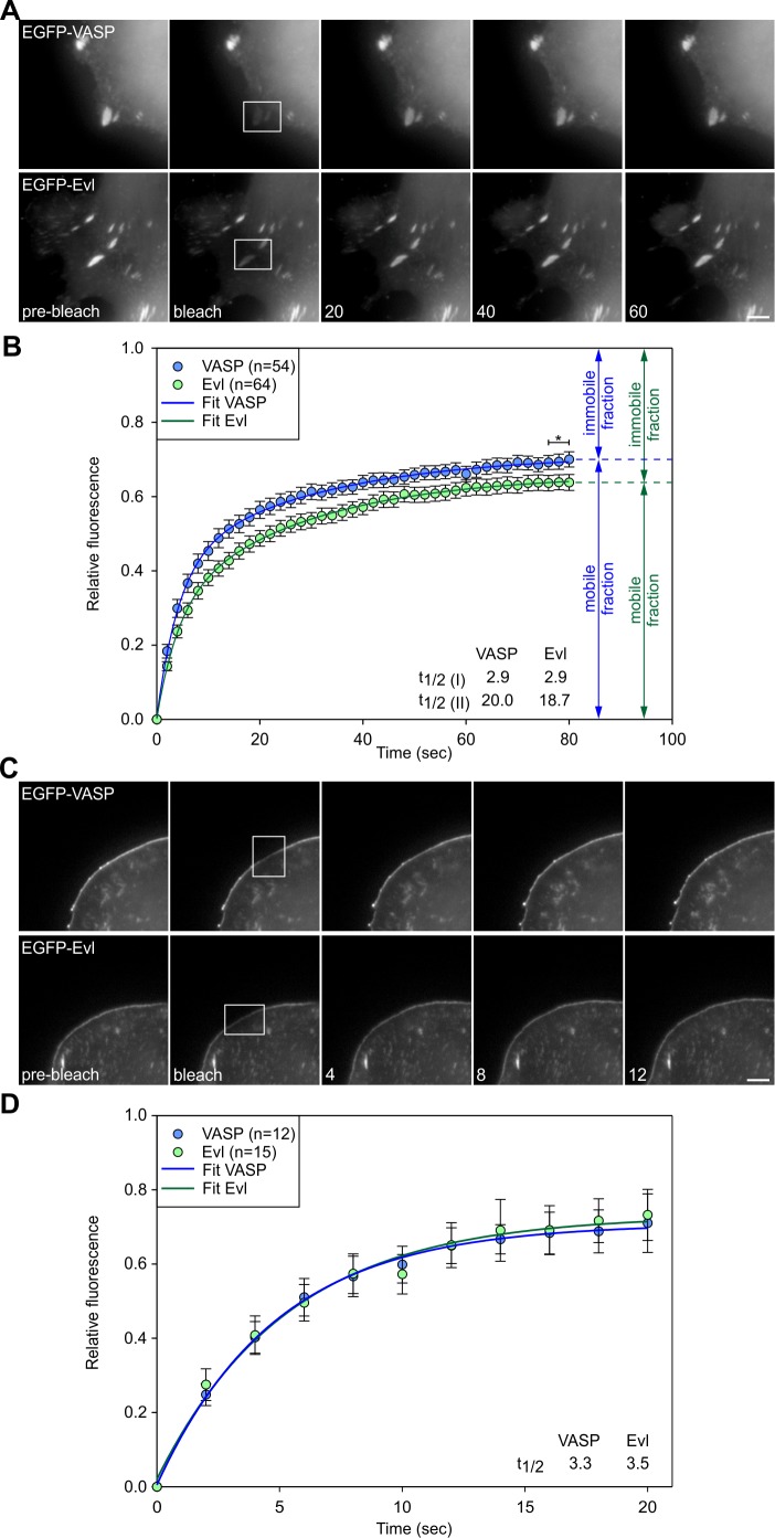
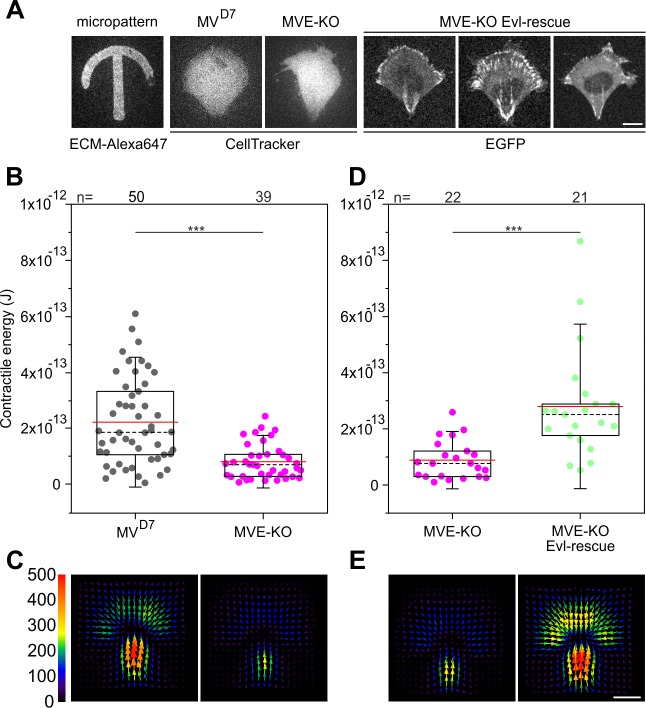
Cell migration entails networks and bundles of actin filaments termed lamellipodia and microspikes or filopodia, respectively, as well as focal adhesions, all of which recruit Ena/VASP family members hitherto thought to antagonize efficient cell motility. However, we find these proteins to act as positive regulators of migration in different murine cell lines. CRISPR/Cas9-mediated loss of Ena/VASP proteins reduced lamellipodial actin assembly and perturbed lamellipodial architecture, as evidenced by changed network geometry as well as reduction of filament length and number that was accompanied by abnormal Arp2/3 complex and heterodimeric capping protein accumulation. Loss of Ena/VASP function also abolished the formation of microspikes normally embedded in lamellipodia, but not of filopodia capable of emanating without lamellipodia. Ena/VASP-deficiency also impaired integrin-mediated adhesion accompanied by reduced traction forces exerted through these structures. Our data thus uncover novel Ena/VASP functions of these actin polymerases that are fully consistent with their promotion of cell migration.

摘要

细胞迁移需要网络和束状肌动蛋白丝,分别称为片状伪足和微刺或丝状伪足,以及粘着斑,所有这些都招募了以前被认为拮抗有效细胞运动的 Ena/VASP 家族成员。然而,我们发现这些蛋白质在不同的小鼠细胞系中作为迁移的正调节剂发挥作用。CRISPR/Cas9 介导的 Ena/VASP 蛋白缺失减少了片状伪足的肌动蛋白组装,并扰乱了片状伪足的结构,这表现在网络几何形状的改变,以及纤维长度和数量的减少,伴随着异常的 Arp2/3 复合物和异二聚体盖帽蛋白的积累。Ena/VASP 功能的丧失也取消了通常嵌入片状伪足的微刺的形成,但不能形成没有片状伪足的丝状伪足。Ena/VASP 缺陷也损害了整合素介导的粘附,同时通过这些结构施加的牵引力减小。因此,我们的数据揭示了这些肌动蛋白聚合酶的新型 Ena/VASP 功能,这完全符合它们促进细胞迁移的作用。

https://cdn.ncbi.nlm.nih.gov/pmc/blobs/258e/7239657/6e9aaaad286c/elife-55351-fig8-figsupp1.jpg
https://cdn.ncbi.nlm.nih.gov/pmc/blobs/258e/7239657/b09b2aa28292/elife-55351-fig1.jpg
https://cdn.ncbi.nlm.nih.gov/pmc/blobs/258e/7239657/8c4490842fb1/elife-55351-fig1-figsupp1.jpg
https://cdn.ncbi.nlm.nih.gov/pmc/blobs/258e/7239657/c1dbed389a0e/elife-55351-fig1-figsupp2.jpg
https://cdn.ncbi.nlm.nih.gov/pmc/blobs/258e/7239657/3f168e176e30/elife-55351-fig1-figsupp3.jpg
https://cdn.ncbi.nlm.nih.gov/pmc/blobs/258e/7239657/ee8216b125ca/elife-55351-fig2.jpg
https://cdn.ncbi.nlm.nih.gov/pmc/blobs/258e/7239657/3cc61d6231f0/elife-55351-fig2-figsupp1.jpg
https://cdn.ncbi.nlm.nih.gov/pmc/blobs/258e/7239657/e319d4a2b744/elife-55351-fig3.jpg
https://cdn.ncbi.nlm.nih.gov/pmc/blobs/258e/7239657/da14b01cc3e4/elife-55351-fig3-figsupp1.jpg
https://cdn.ncbi.nlm.nih.gov/pmc/blobs/258e/7239657/d4763581bb83/elife-55351-fig4.jpg
https://cdn.ncbi.nlm.nih.gov/pmc/blobs/258e/7239657/5bd75420f7f6/elife-55351-fig4-figsupp1.jpg
https://cdn.ncbi.nlm.nih.gov/pmc/blobs/258e/7239657/615388762869/elife-55351-fig4-figsupp2.jpg
https://cdn.ncbi.nlm.nih.gov/pmc/blobs/258e/7239657/3fa635702005/elife-55351-fig5.jpg
https://cdn.ncbi.nlm.nih.gov/pmc/blobs/258e/7239657/2fb9ea20a11d/elife-55351-fig5-figsupp1.jpg
https://cdn.ncbi.nlm.nih.gov/pmc/blobs/258e/7239657/aca4e83f8d1e/elife-55351-fig6.jpg
https://cdn.ncbi.nlm.nih.gov/pmc/blobs/258e/7239657/d2224aa3a8d6/elife-55351-fig7.jpg
https://cdn.ncbi.nlm.nih.gov/pmc/blobs/258e/7239657/9e6a6edbd80f/elife-55351-fig7-figsupp1.jpg
https://cdn.ncbi.nlm.nih.gov/pmc/blobs/258e/7239657/4521f9f038c3/elife-55351-fig7-figsupp2.jpg
https://cdn.ncbi.nlm.nih.gov/pmc/blobs/258e/7239657/8bac4a721534/elife-55351-fig8.jpg
https://cdn.ncbi.nlm.nih.gov/pmc/blobs/258e/7239657/6e9aaaad286c/elife-55351-fig8-figsupp1.jpg

相似文献

1
Loss of Ena/VASP interferes with lamellipodium architecture, motility and integrin-dependent adhesion.Ena/VASP 的缺失会干扰片状伪足的结构、运动和整合素依赖性黏附。
Elife. 2020 May 11;9:e55351. doi: 10.7554/eLife.55351.
2
Antagonism between Ena/VASP proteins and actin filament capping regulates fibroblast motility.Ena/VASP蛋白与肌动蛋白丝封端之间的拮抗作用调节成纤维细胞的运动。
Cell. 2002 May 17;109(4):509-21. doi: 10.1016/s0092-8674(02)00731-6.
3
Reconstitution of the transition from a lamellipodia- to filopodia-like actin network with purified proteins.用纯化蛋白重构从片状伪足到丝状伪足样肌动蛋白网络的转变。
Eur J Cell Biol. 2023 Dec;102(4):151367. doi: 10.1016/j.ejcb.2023.151367. Epub 2023 Oct 20.
4
Ena/VASP clustering at microspike tips involves lamellipodin but not I-BAR proteins, and absolutely requires unconventional myosin-X.微刺尖端的 Ena/VASP 聚集涉及片状蛋白但不涉及 I-BAR 蛋白,绝对需要非典型肌球蛋白-X。
Proc Natl Acad Sci U S A. 2023 Jan 10;120(2):e2217437120. doi: 10.1073/pnas.2217437120. Epub 2023 Jan 4.
5
Efficiency of lamellipodia protrusion is determined by the extent of cytosolic actin assembly.片状伪足突出的效率取决于胞质肌动蛋白组装的程度。
Mol Biol Cell. 2017 May 15;28(10):1311-1325. doi: 10.1091/mbc.E16-05-0334. Epub 2017 Mar 22.
6
Ena/VASP proteins have an anti-capping independent function in filopodia formation.埃娜/血管成形素相关蛋白(Ena/VASP)在丝状伪足形成中具有一种独立于帽化作用的功能。
Mol Biol Cell. 2007 Jul;18(7):2579-91. doi: 10.1091/mbc.e06-11-0990. Epub 2007 May 2.
7
Capping protein is dispensable for polarized actin network growth and actin-based motility.衔接蛋白对于极化肌动蛋白网络的生长和基于肌动蛋白的运动是可有可无的。
J Biol Chem. 2020 Nov 6;295(45):15366-15375. doi: 10.1074/jbc.RA120.015009. Epub 2020 Aug 31.
8
Ena/VASP proteins can regulate distinct modes of actin organization at cadherin-adhesive contacts.埃娜/血管舒缩刺激蛋白(Ena/VASP)家族蛋白可在钙黏蛋白黏附连接处调节肌动蛋白组装的不同模式。
Mol Biol Cell. 2006 Mar;17(3):1085-95. doi: 10.1091/mbc.e05-07-0644. Epub 2005 Dec 21.
9
WAVE binds Ena/VASP for enhanced Arp2/3 complex-based actin assembly.WAVE与Ena/VASP结合,以增强基于Arp2/3复合物的肌动蛋白组装。
Mol Biol Cell. 2015 Jan 1;26(1):55-65. doi: 10.1091/mbc.E14-07-1200. Epub 2014 Oct 29.
10
Lamellipodin tunes cell migration by stabilizing protrusions and promoting adhesion formation.薄束蛋白通过稳定突起和促进黏附形成来调节细胞迁移。
J Cell Sci. 2020 Apr 9;133(7):jcs239020. doi: 10.1242/jcs.239020.

引用本文的文献

1
Rnd3 deletion affects neuroblast behavior through the RhoA/ROCK pathway but not neural stem cells in postnatal mice subventricular zone.Rnd3缺失通过RhoA/ROCK信号通路影响产后小鼠脑室下区神经母细胞的行为,但不影响神经干细胞。
Front Cell Dev Biol. 2025 Jun 23;13:1612177. doi: 10.3389/fcell.2025.1612177. eCollection 2025.
2
Probing the protrusions: lamellipodia and filopodia in cancer invasion and beyond.探索突起结构:板状伪足和丝状伪足在癌症侵袭及其他方面的作用
Mechanobiol Med. 2024 Mar 20;2(2):100064. doi: 10.1016/j.mbm.2024.100064. eCollection 2024 Jun.
3
The NHSL1-A complex interacts with the Arp2/3 complex and controls cell migration efficiency and chemotaxis.

本文引用的文献

1
Mechanosensing during directed cell migration requires dynamic actin polymerization at focal adhesions.在定向细胞迁移过程中,机械感觉需要在黏着斑处进行动态肌动蛋白聚合。
J Cell Biol. 2019 Dec 2;218(12):4215-4235. doi: 10.1083/jcb.201902101. Epub 2019 Oct 8.
2
Distinct Interaction Sites of Rac GTPase with WAVE Regulatory Complex Have Non-redundant Functions in Vivo.Rac GTPase 与 WAVE 调节复合物的独特相互作用位点在体内具有非冗余的功能。
Curr Biol. 2018 Nov 19;28(22):3674-3684.e6. doi: 10.1016/j.cub.2018.10.002. Epub 2018 Nov 1.
3
Roles for Ena/VASP proteins in FMNL3-mediated filopodial assembly.
NHSL1-A复合物与Arp2/3复合物相互作用,并控制细胞迁移效率和趋化性。
bioRxiv. 2025 Mar 13:2025.03.13.643034. doi: 10.1101/2025.03.13.643034.
4
The PTTG1/VASP axis promotes oral squamous cell carcinoma metastasis by modulating focal adhesion and actin filaments.PTTG1/VASP轴通过调节粘着斑和肌动蛋白丝促进口腔鳞状细胞癌转移。
Mol Oncol. 2025 May;19(5):1517-1531. doi: 10.1002/1878-0261.13779. Epub 2025 Jan 10.
5
Sequestration of ribosomal subunits as inactive 80S by targeting eIF6 limits mitotic exit and cancer progression.通过靶向真核起始因子6(eIF6)将核糖体亚基隔离为无活性的80S核糖体,会限制有丝分裂退出和癌症进展。
Nucleic Acids Res. 2025 Feb 8;53(4). doi: 10.1093/nar/gkae1272.
6
An allosteric inhibitor of RhoGAP class-IX myosins suppresses the metastatic features of cancer cells.一种变构 RhoGAP 类肌球蛋白的抑制剂抑制了癌细胞的转移特征。
Nat Commun. 2024 Nov 16;15(1):9947. doi: 10.1038/s41467-024-54181-6.
7
Decisive role of mDia-family formins in cell cortex function of highly adherent cells.mDia 家族formin 在高黏附细胞细胞皮层功能中的决定性作用。
Sci Adv. 2024 Nov;10(44):eadp5929. doi: 10.1126/sciadv.adp5929. Epub 2024 Oct 30.
8
Membrane Ruffles: Composition, Function, Formation and Visualization.膜皱襞:组成、功能、形成和可视化。
Int J Mol Sci. 2024 Oct 12;25(20):10971. doi: 10.3390/ijms252010971.
9
Ancient eukaryotic protein interactions illuminate modern genetic traits and disorders.古代真核生物蛋白质相互作用揭示现代遗传特征和疾病。
bioRxiv. 2024 May 29:2024.05.26.595818. doi: 10.1101/2024.05.26.595818.
10
Decreased Tiam1-mediated Rac1 activation is responsible for impaired directional persistence of chondrocyte migration in microtia.Tiam1 介导的 Rac1 活化减少导致小耳畸形中软骨细胞迁移的定向持续性受损。
J Cell Mol Med. 2024 Jun;28(11):e18443. doi: 10.1111/jcmm.18443.
Ena/VASP 蛋白在 FMNL3 介导的丝状伪足组装中的作用。
J Cell Sci. 2018 Oct 29;131(21):jcs220814. doi: 10.1242/jcs.220814.
4
Focal Adhesions Undergo Longitudinal Splitting into Fixed-Width Units.焦点黏附会纵向分裂成固定宽度的单位。
Curr Biol. 2018 Jul 9;28(13):2033-2045.e5. doi: 10.1016/j.cub.2018.04.073. Epub 2018 Jun 14.
5
Micromanipulation Techniques Allowing Analysis of Morphogenetic Dynamics and Turnover of Cytoskeletal Regulators.允许分析形态发生动力学和细胞骨架调节因子周转的显微操作技术
J Vis Exp. 2018 May 12(135):57643. doi: 10.3791/57643.
6
Analysis of Random Migration of Dictyostelium Amoeba in Confined and Unconfined Environments.盘基网柄菌变形虫在受限和非受限环境中的随机迁移分析
Methods Mol Biol. 2018;1749:341-350. doi: 10.1007/978-1-4939-7701-7_24.
7
The Arp2/3 Regulatory System and Its Deregulation in Cancer.Arp2/3 调控系统及其在癌症中的失调。
Physiol Rev. 2018 Jan 1;98(1):215-238. doi: 10.1152/physrev.00006.2017.
8
Cortactin: Cell Functions of A Multifaceted Actin-Binding Protein.细胞黏着蛋白:多功能肌动蛋白结合蛋白的细胞功能。
Trends Cell Biol. 2018 Feb;28(2):79-98. doi: 10.1016/j.tcb.2017.10.009. Epub 2017 Nov 20.
9
Glyoxal as an alternative fixative to formaldehyde in immunostaining and super-resolution microscopy.乙二醛作为免疫染色和超分辨率显微镜检查中甲醛的替代固定剂。
EMBO J. 2018 Jan 4;37(1):139-159. doi: 10.15252/embj.201695709. Epub 2017 Nov 16.
10
WH2 and proline-rich domains of WASP-family proteins collaborate to accelerate actin filament elongation.WH2 和富含脯氨酸的结构域的 WASP 家族蛋白协同作用加速肌动蛋白丝的延伸。
EMBO J. 2018 Jan 4;37(1):102-121. doi: 10.15252/embj.201797039. Epub 2017 Nov 15.