Nelson Amy R, Sagare Meghana A, Wang Yaoming, Kisler Kassandra, Zhao Zhen, Zlokovic Berislav V
Department of Physiology and Neuroscience, Zilkha Neurogenetic Institute, University of Southern California, Los Angeles, CA, United States.
Front Aging Neurosci. 2020 Apr 29;12:108. doi: 10.3389/fnagi.2020.00108. eCollection 2020.
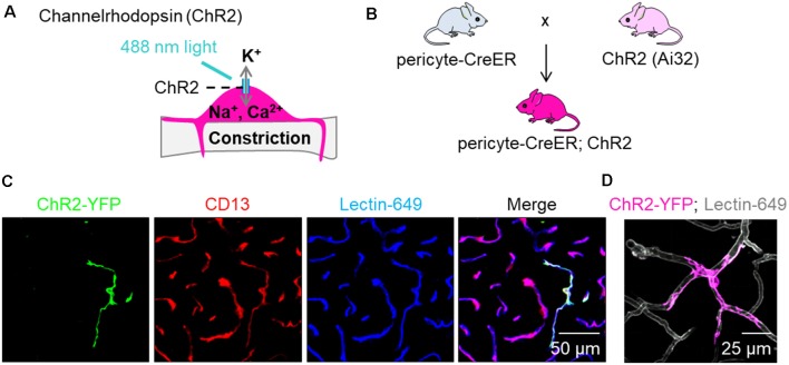
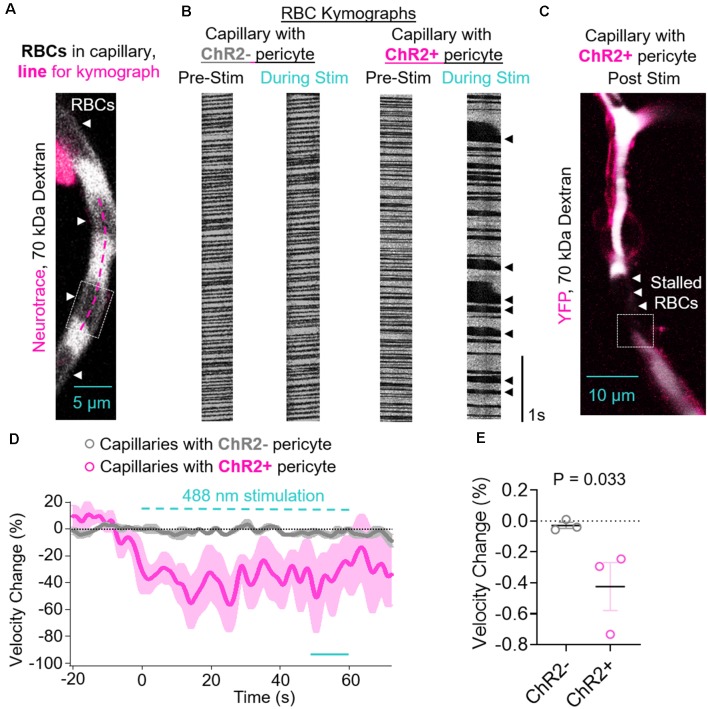
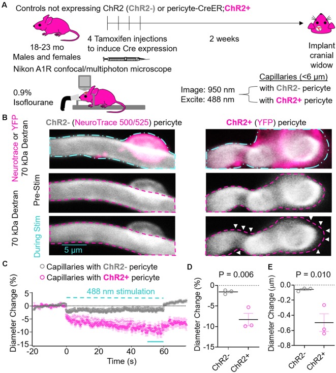
Brains depend on blood flow for the delivery of oxygen and nutrients essential for proper neuronal and synaptic functioning. French physiologist Rouget was the first to describe pericytes in 1873 as regularly arranged longitudinal amoeboid cells on capillaries that have a muscular coat, implying that these are contractile cells that regulate blood flow. Although there have been >30 publications from different groups, including our group, demonstrating that pericytes are contractile cells that can regulate hemodynamic responses in the brain, the role of pericytes in controlling cerebral blood flow (CBF) has not been confirmed by all studies. Moreover, recent studies using different optogenetic models to express light-sensitive channelrhodopsin-2 (ChR2) cation channels in pericytes were not conclusive; one, suggesting that pericytes expressing ChR2 do not contract after light stimulus, and the other, demonstrating contraction of pericytes expressing ChR2 after light stimulus. Since two-photon optogenetics provides a powerful tool to study mechanisms of blood flow regulation at the level of brain capillaries, we re-examined the contractility of brain pericytes using a new optogenetic model developed by crossing our new inducible pericyte-specific CreER mouse line with ChR2 mice. We induced expression of ChR2 in pericytes with tamoxifen, excited ChR2 by 488 nm light, and monitored pericyte contractility, brain capillary diameter changes, and red blood cell (RBC) velocity in aged mice by two-photon microscopy. Excitation of ChR2 resulted in pericyte contraction followed by constriction of the underlying capillary leading to approximately an 8% decrease ( = 0.006) in capillary diameter. ChR2 excitation in pericytes substantially reduced capillary RBC flow by 42% ( = 0.03) during the stimulation period compared to the velocity before stimulation. Our data suggests that pericytes contract and regulate capillary blood flow in the aging mouse brain. By extension, this might have implications for neurological disorders of the aging human brain associated with neurovascular dysfunction and pericyte loss such as stroke and Alzheimer's disease.
大脑依赖血液流动来输送对神经元和突触正常功能至关重要的氧气和营养物质。1873年,法国生理学家鲁热首次将周细胞描述为毛细血管上规则排列的纵向阿米巴样细胞,这些毛细血管有一层肌肉层,这意味着它们是调节血流的收缩细胞。尽管包括我们小组在内的不同研究团队已发表了30多篇论文,证明周细胞是可调节大脑血流动力学反应的收缩细胞,但并非所有研究都证实了周细胞在控制脑血流量(CBF)中的作用。此外,最近使用不同光遗传学模型在周细胞中表达光敏感通道视紫红质-2(ChR2)阳离子通道的研究结果并不确凿;一项研究表明,表达ChR2的周细胞在光刺激后不会收缩,而另一项研究则表明,表达ChR2的周细胞在光刺激后会收缩。由于双光子光遗传学为研究脑毛细血管水平的血流调节机制提供了一个强大的工具,我们使用一种新的光遗传学模型重新研究了脑周细胞的收缩性,该模型是通过将我们新的可诱导周细胞特异性CreER小鼠品系与ChR2小鼠杂交而开发的。我们用他莫昔芬诱导周细胞中ChR2的表达,用488nm光激发ChR2,并通过双光子显微镜监测老年小鼠的周细胞收缩性、脑毛细血管直径变化和红细胞(RBC)速度。ChR2的激发导致周细胞收缩,随后下层毛细血管收缩,导致毛细血管直径大约减少8%(P = 0.006)。与刺激前的速度相比,在刺激期间,周细胞中ChR2的激发使毛细血管RBC流量大幅减少了42%(P = 0.03)。我们的数据表明,周细胞在衰老小鼠大脑中收缩并调节毛细血管血流。由此推断,这可能对与神经血管功能障碍和周细胞丢失相关的衰老人类大脑神经疾病(如中风和阿尔茨海默病)具有重要意义。