Matono Rumi, Ninomiya Mizuki, Morita Kazutoyo, Tomino Takahiro, Oshiro Yumi, Yokota Tomoyuki, Nishizaki Takashi
Department of Surgery, Matsuyama Red Cross Hospital, 1 Bunkyo-cho, Matsuyama City, Ehime, 790-8524, Japan.
Department of Diagnostic Pathology, Matsuyama Red Cross Hospital, Matsuyama City, Ehime, Japan.
Surg Case Rep. 2020 May 15;6(1):103. doi: 10.1186/s40792-020-00864-3.
Intraductal papillary neoplasm of the bile duct (IPNB) is characterized by an intraluminal, growing papillary tumor covered by neoplastic biliary epithelial cells with a fine fibrovascular core. IPNB was introduced as a precancerous and early neoplastic lesion in the 2010 World Health Organization classification of tumors of the digestive system. IPNB eventually invades the bile duct wall and progresses to invasive cholangiocarcinoma. IPNB resembles intraductal papillary mucinous neoplasm of the pancreas (IPMN), particularly the main pancreatic duct type. IPNB cases, possibly corresponding to branch-type IPMN, have been recently reported, and these cases involved the peribiliary glands significantly and showed gross cystic dilatation. Small branch-type intrahepatic IPNB often mimics simple liver cysts, making the diagnosis of IPNB difficult. Some literature recommended surgical resection for treatment. Laparoscopic resection is a good treatment option for small tumor. We herein present the case of branch-type IPNB that was treated with laparoscopic anatomical liver resection 5 years after being detected.
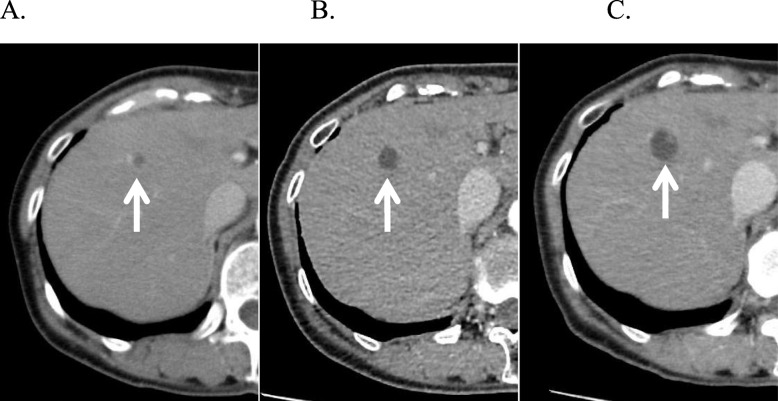
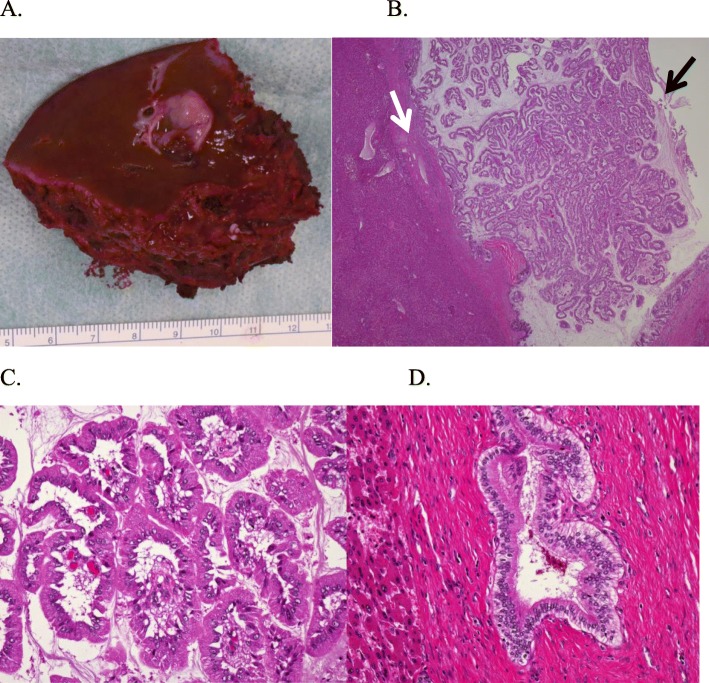
A 64-year-old woman was undergoing follow-up for primary aldosteronism. In 2012, follow-up computed tomography (CT) incidentally revealed a 7-mm cystic lesion in segment 8 of the liver. From 2012 to 2017, the cystic lesion kept increasing in size, reaching 17 mm. In 2017, CT also revealed a 13-mm mural nodule in the cyst wall. Therefore, the patient was referred to our department for possible malignancy. We suspected a branch-type IPNB; however, the mass was small and diagnosis could not be made without performing biopsy. Accordingly, surgical resection was performed for diagnosis and treatment. Because branch-type IPNB might show horizontal spread through the intrahepatic bile duct, we believed that anatomical resection of the liver was appropriate considering the malignant potential of the lesion. Therefore, laparoscopic anatomical resection of segment 8 of the liver was performed. The resected tumor measured 17 mm and was histologically diagnosed as a high-grade IPNB.
Branch-type IPNBs are rare but can potentially lead to malignant tumors. Surgical resection is the treatment of choice, with laparoscopic anatomical resection being a good treatment option for this small tumor.
胆管内乳头状肿瘤(IPNB)的特征是管腔内生长的乳头状肿瘤,表面覆盖有肿瘤性胆管上皮细胞,有纤细的纤维血管轴心。IPNB在2010年世界卫生组织消化系统肿瘤分类中被列为一种癌前和早期肿瘤性病变。IPNB最终会侵犯胆管壁并进展为浸润性胆管癌。IPNB类似于胰腺导管内乳头状黏液性肿瘤(IPMN),尤其是主胰管型。最近有报道称存在可能对应于分支型IPMN的IPNB病例,这些病例显著累及胆管周围腺体并表现为肉眼可见的囊状扩张。小分支型肝内IPNB常类似单纯肝囊肿,使得IPNB的诊断困难。一些文献推荐手术切除作为治疗方法。腹腔镜切除对于小肿瘤是一个很好的治疗选择。我们在此报告一例分支型IPNB病例,该病例在被发现5年后接受了腹腔镜解剖性肝切除术治疗。
一名64岁女性因原发性醛固酮增多症正在接受随访。2012年,随访计算机断层扫描(CT)偶然发现肝脏第8段有一个7毫米的囊性病变。从2012年到2017年,该囊性病变大小持续增大,达到17毫米。2017年,CT还显示囊肿壁上有一个13毫米的壁结节。因此,该患者被转诊至我科以排查是否存在恶性病变。我们怀疑是分支型IPNB;然而,肿块较小,不进行活检无法确诊。因此,为了诊断和治疗进行了手术切除。由于分支型IPNB可能通过肝内胆管出现横向扩散,考虑到病变的恶性潜能,我们认为进行肝脏解剖性切除是合适的。因此,实施了肝脏第8段的腹腔镜解剖性切除。切除的肿瘤大小为17毫米,组织学诊断为高级别IPNB。
分支型IPNB罕见,但有可能发展为恶性肿瘤。手术切除是首选治疗方法,腹腔镜解剖性切除对于这种小肿瘤是一个很好的治疗选择。