• 文献检索
  • 文档翻译
  • 深度研究
  • 学术资讯
  • Suppr Zotero 插件Zotero 插件
  • 邀请有礼
  • 套餐&价格
  • 历史记录
应用&插件
Suppr Zotero 插件Zotero 插件浏览器插件Mac 客户端Windows 客户端微信小程序
定价
高级版会员购买积分包购买API积分包
服务
文献检索文档翻译深度研究API 文档MCP 服务
关于我们
关于 Suppr公司介绍联系我们用户协议隐私条款
关注我们

Suppr 超能文献

核心技术专利:CN118964589B侵权必究
粤ICP备2023148730 号-1Suppr @ 2026

文献检索

告别复杂PubMed语法,用中文像聊天一样搜索,搜遍4000万医学文献。AI智能推荐,让科研检索更轻松。

立即免费搜索

文件翻译

保留排版,准确专业,支持PDF/Word/PPT等文件格式,支持 12+语言互译。

免费翻译文档

深度研究

AI帮你快速写综述,25分钟生成高质量综述,智能提取关键信息,辅助科研写作。

立即免费体验

间质、血管和小管周围层中表达干细胞性 Leydig 细胞标志物的人类成年睾丸细胞的比较分析。

A comparative analysis of human adult testicular cells expressing stem Leydig cell markers in the interstitium, vasculature, and peritubular layer.

机构信息

Center for Reproductive Medicine, Amsterdam Reproduction and Development, Amsterdam UMC, University of Amsterdam, Amsterdam, The Netherlands.

Department of Animal Sciences, Human and Animal Physiology, Wageningen University, Wageningen, The Netherlands.

出版信息

Andrology. 2020 Sep;8(5):1265-1276. doi: 10.1111/andr.12817. Epub 2020 Jun 1.

DOI:10.1111/andr.12817
PMID:32416031
原文链接:https://pmc.ncbi.nlm.nih.gov/articles/PMC7496384/
Abstract

BACKGROUND

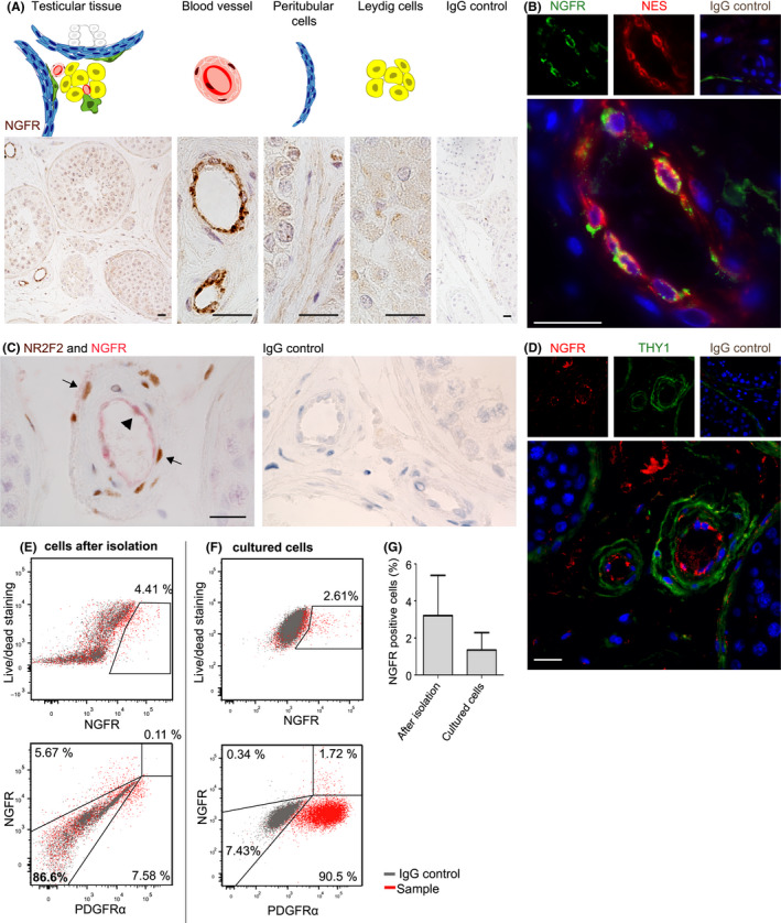
Origin of human adult Leydig cells (ALCs) is not well understood. This might be partly due to limited data available on the identification and location of human precursor and stem Leydig cells (SLCs) which hampers the study on the development of ALCs.

OBJECTIVES

The aim of the present study was to investigate whether described human (PDGFRα, NGFR) and rodent (NES, PDGFRα, THY1, NR2F2) SLC markers are expressed by a common cell population within human adult testicular interstitial cells in vivo and before and after in vitro propagation.

MATERIALS AND METHODS

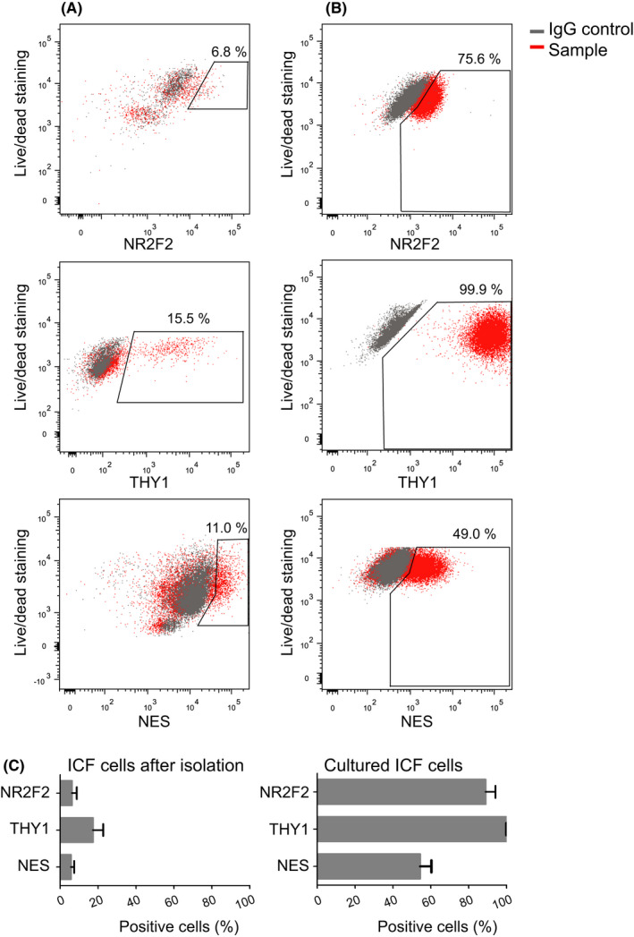
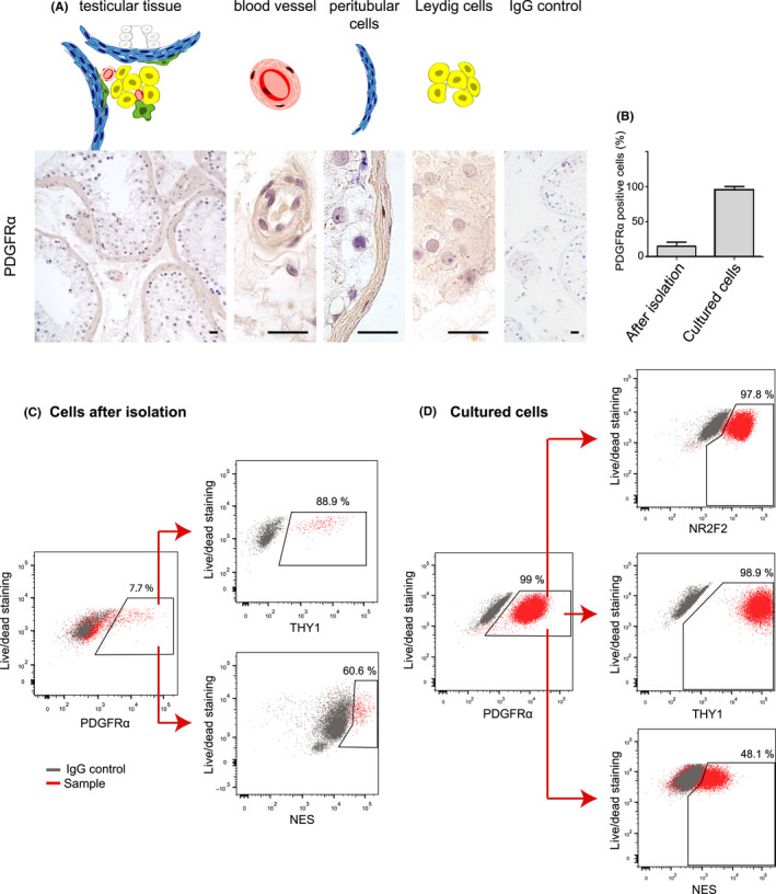
Immunohistochemical analyses were used to identify localization of human adult testicular interstitial cells expressing described SLC markers. Next, interstitial cells were isolated and cultured. The percentage of cells expressing one or more SLC markers was determined before and after culture using flow cytometry.

RESULTS

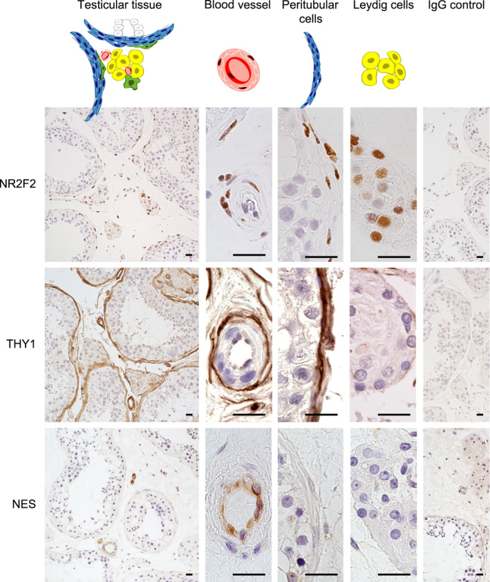
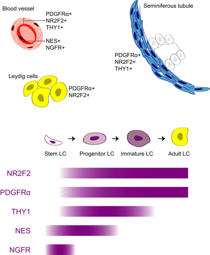
NR2F2 and PDGFRα were present in peritubular, perivascular, and Leydig cells, while THY1 was expressed in peritubular and perivascular cells. Although NES and NGFR were expressed in endothelial cells, co-localization with PDGFRα was found for both in vitro, although for NGFR only after culture. All marker positive cells were able to undergo propagation in vitro.

DISCUSSION

The partly overlap in localization and overlap in expression in human testicular cells indicate that PDGFRα, NR2F2, and THY1 are expressed within the same ALC developmental lineage from SLCs. Based on the in vitro results, this is also true for NES and after in vitro propagation for NGFR.

CONCLUSION

Our results that earlier described SLC markers are expressed in overlapping human interstitial cell population opens up further research strategies aiming for a better insight in the Leydig cell lineage and will be helpful for development of strategies to cure ALC dysfunction.

摘要

背景

人类成年莱迪希细胞(ALC)的起源尚不清楚。这可能部分归因于目前关于人类前体和干细胞莱迪希细胞(SLC)的鉴定和定位的可用数据有限,这阻碍了对 ALC 发育的研究。

目的

本研究旨在探讨已描述的人类(PDGFRα、NGFR)和啮齿动物(NES、PDGFRα、THY1、NR2F2)SLC 标志物是否在体内和体外增殖前后表达于人类成年睾丸间质细胞中的共同细胞群。

材料和方法

免疫组织化学分析用于鉴定表达已描述的 SLC 标志物的人类成年睾丸间质细胞的定位。然后,分离和培养间质细胞。使用流式细胞术在培养前后确定表达一种或多种 SLC 标志物的细胞的百分比。

结果

NR2F2 和 PDGFRα 存在于小管周围、血管周围和莱迪希细胞中,而 THY1 则存在于小管周围和血管周围细胞中。尽管 NES 和 NGFR 存在于内皮细胞中,但在体外发现两者与 PDGFRα 共表达,尽管 NGFR 仅在培养后才表达。所有标记阳性细胞均能够在体外增殖。

讨论

在人类睾丸细胞中的定位部分重叠和表达重叠表明,PDGFRα、NR2F2 和 THY1 是在 SLC 内的相同 ALC 发育谱系中表达的。基于体外结果,这对于 NES 也是如此,并且对于体外培养后的 NGFR 也是如此。

结论

我们的结果表明,先前描述的 SLC 标志物在重叠的人类间质细胞群中表达,这为进一步研究莱迪希细胞谱系提供了更多的研究策略,并有助于开发治疗 ALC 功能障碍的策略。

https://cdn.ncbi.nlm.nih.gov/pmc/blobs/a05f/7496384/4aa6521ced83/ANDR-8-1265-g005.jpg
https://cdn.ncbi.nlm.nih.gov/pmc/blobs/a05f/7496384/742746e3f254/ANDR-8-1265-g001.jpg
https://cdn.ncbi.nlm.nih.gov/pmc/blobs/a05f/7496384/0639db170aa4/ANDR-8-1265-g002.jpg
https://cdn.ncbi.nlm.nih.gov/pmc/blobs/a05f/7496384/da56895ebc4b/ANDR-8-1265-g003.jpg
https://cdn.ncbi.nlm.nih.gov/pmc/blobs/a05f/7496384/d76ac6fd551a/ANDR-8-1265-g004.jpg
https://cdn.ncbi.nlm.nih.gov/pmc/blobs/a05f/7496384/4aa6521ced83/ANDR-8-1265-g005.jpg

相似文献

1
A comparative analysis of human adult testicular cells expressing stem Leydig cell markers in the interstitium, vasculature, and peritubular layer.间质、血管和小管周围层中表达干细胞性 Leydig 细胞标志物的人类成年睾丸细胞的比较分析。
Andrology. 2020 Sep;8(5):1265-1276. doi: 10.1111/andr.12817. Epub 2020 Jun 1.
2
Primary human testicular PDGFRα+ cells are multipotent and can be differentiated into cells with Leydig cell characteristics in vitro.原代人睾丸 PDGFRα+ 细胞具有多能性,并且可以在体外分化为具有间质细胞特征的细胞。
Hum Reprod. 2019 Sep 29;34(9):1621-1631. doi: 10.1093/humrep/dez131.
3
Endosialin defines human stem Leydig cells with regenerative potential.内唾液酸糖蛋白(endosialin)可鉴定具有再生潜能的人源睾丸间质干细胞。
Hum Reprod. 2020 Oct 1;35(10):2197-2212. doi: 10.1093/humrep/deaa174.
4
Leydig cell stem cells: Identification, proliferation and differentiation.睾丸间质干细胞:鉴定、增殖与分化
Mol Cell Endocrinol. 2017 Apr 15;445:65-73. doi: 10.1016/j.mce.2016.10.010. Epub 2016 Oct 12.
5
Characterization of Nestin-positive stem Leydig cells as a potential source for the treatment of testicular Leydig cell dysfunction.巢蛋白阳性的睾丸间质干细胞特性研究:一种治疗睾丸间质细胞功能障碍的潜在细胞来源
Cell Res. 2014 Dec;24(12):1466-85. doi: 10.1038/cr.2014.149. Epub 2014 Nov 21.
6
Human testicular peritubular cells host putative stem Leydig cells with steroidogenic capacity.人类睾丸的睾丸间质细胞含有具有类固醇生成能力的假定干细胞样睾丸间质细胞。
J Clin Endocrinol Metab. 2014 Jul;99(7):E1227-35. doi: 10.1210/jc.2013-4199. Epub 2014 Mar 31.
7
Stem Leydig Cells in the Adult Testis: Characterization, Regulation and Potential Applications.成年睾丸中的精原干细胞:特征、调控及潜在应用。
Endocr Rev. 2020 Feb 1;41(1):22-32. doi: 10.1210/endrev/bnz013.
8
Expression patterns of DLK1 and INSL3 identify stages of Leydig cell differentiation during normal development and in testicular pathologies, including testicular cancer and Klinefelter syndrome.DLK1和INSL3的表达模式可确定正常发育过程中以及包括睾丸癌和克氏综合征在内的睾丸病理状态下睾丸间质细胞分化的阶段。
Hum Reprod. 2014 Aug;29(8):1637-50. doi: 10.1093/humrep/deu124. Epub 2014 Jun 7.
9
Transplantation of CD51 Stem Leydig Cells: A New Strategy for the Treatment of Testosterone Deficiency.CD51 干细胞睾丸间质细胞移植:治疗睾酮缺乏症的新策略。
Stem Cells. 2017 May;35(5):1222-1232. doi: 10.1002/stem.2569. Epub 2017 Mar 5.
10
Characterization and differentiation of CD51 Stem Leydig cells in adult mouse testes.鉴定和分化成年小鼠睾丸中的 CD51 干细胞性 Leydig 细胞。
Mol Cell Endocrinol. 2019 Aug 1;493:110449. doi: 10.1016/j.mce.2019.110449. Epub 2019 May 15.

引用本文的文献

1
Identification and isolation of human testicular peritubular myoid cells and Leydig cells by a combination of ITGA9 and NGFR.通过整合素α9(ITGA9)和神经生长因子受体(NGFR)组合鉴定和分离人睾丸周细胞和睾丸间质细胞。
Reprod Biol Endocrinol. 2025 May 31;23(1):82. doi: 10.1186/s12958-025-01389-w.
2
Roles of PDGF/PDGFR signaling in various organs.血小板衍生生长因子/血小板衍生生长因子受体信号通路在各器官中的作用。
Korean J Physiol Pharmacol. 2025 Mar 1;29(2):139-155. doi: 10.4196/kjpp.24.309. Epub 2024 Oct 31.
3
Effects of Leydig cell elimination on testicular interstitial cell populations: characterization by scRNA-seq and immunocytochemical techniques.

本文引用的文献

1
The Dynamic Transcriptional Cell Atlas of Testis Development during Human Puberty.人类青春期睾丸发育的动态转录细胞图谱。
Cell Stem Cell. 2020 Feb 6;26(2):262-276.e4. doi: 10.1016/j.stem.2019.12.005. Epub 2020 Jan 9.
2
Insights into replicative senescence of human testicular peritubular cells.人睾丸小管周细胞复制性衰老的研究进展。
Sci Rep. 2019 Oct 21;9(1):15052. doi: 10.1038/s41598-019-51380-w.
3
Primary human testicular PDGFRα+ cells are multipotent and can be differentiated into cells with Leydig cell characteristics in vitro.
Leydig 细胞消除对睾丸间质细胞群的影响:通过 scRNA-seq 和免疫细胞化学技术进行表征。
Front Endocrinol (Lausanne). 2024 Aug 20;15:1423801. doi: 10.3389/fendo.2024.1423801. eCollection 2024.
4
WNT5A regulates the proliferation, apoptosis and stemness of human stem Leydig cells via the β-catenin signaling pathway.WNT5A 通过β-catenin 信号通路调节人源性精原干细胞的增殖、凋亡和干性。
Cell Mol Life Sci. 2024 Feb 17;81(1):93. doi: 10.1007/s00018-023-05077-z.
5
Nuclear Receptor Gene Variants Underlying Disorders/Differences of Sex Development through Abnormal Testicular Development.核受体基因变异与睾丸发育异常导致的性别发育障碍/差异。
Biomolecules. 2023 Apr 19;13(4):691. doi: 10.3390/biom13040691.
6
The Molecular Signature of Human Testicular Peritubular Cells Revealed by Single-Cell Analysis.单细胞分析揭示的人类睾丸支持细胞的分子特征。
Cells. 2022 Nov 19;11(22):3685. doi: 10.3390/cells11223685.
7
Identification of Rat Testicular Leydig Precursor Cells by Single-Cell-RNA-Sequence Analysis.通过单细胞RNA序列分析鉴定大鼠睾丸间质前体细胞
Front Cell Dev Biol. 2022 Feb 15;10:805249. doi: 10.3389/fcell.2022.805249. eCollection 2022.
8
Effects of Midazolam on the Development of Adult Leydig Cells From Stem Cells .咪达唑仑对干细胞来源的成年莱迪希细胞发育的影响。
Front Endocrinol (Lausanne). 2021 Nov 12;12:765251. doi: 10.3389/fendo.2021.765251. eCollection 2021.
9
Insights into the Regulation on Proliferation and Differentiation of Stem Leydig Cells.深入了解调控精原干细胞增殖与分化的机制
Stem Cell Rev Rep. 2021 Oct;17(5):1521-1533. doi: 10.1007/s12015-021-10133-x. Epub 2021 Feb 17.
10
ITGA6+ Human Testicular Cell Populations Acquire a Mesenchymal Rather than Germ Cell Transcriptional Signature during Long-Term Culture.ITGA6+ 人类睾丸细胞群体在长期培养过程中获得间充质而非生殖细胞转录特征。
Int J Mol Sci. 2020 Nov 4;21(21):8269. doi: 10.3390/ijms21218269.
原代人睾丸 PDGFRα+ 细胞具有多能性,并且可以在体外分化为具有间质细胞特征的细胞。
Hum Reprod. 2019 Sep 29;34(9):1621-1631. doi: 10.1093/humrep/dez131.
4
Endothelial to Mesenchymal Transition: Role in Physiology and in the Pathogenesis of Human Diseases.内皮细胞向间充质细胞转化:在生理和人类疾病发病机制中的作用。
Physiol Rev. 2019 Apr 1;99(2):1281-1324. doi: 10.1152/physrev.00021.2018.
5
Fetal Leydig cells dedifferentiate and serve as adult Leydig stem cells.胎儿莱迪希细胞去分化并作为成体莱迪希干细胞发挥作用。
Development. 2018 Dec 5;145(23):dev169136. doi: 10.1242/dev.169136.
6
Transplanted human p75-positive stem Leydig cells replace disrupted Leydig cells for testosterone production.移植的人 p75 阳性干细胞莱迪希细胞替代破坏的莱迪希细胞以产生睾酮。
Cell Death Dis. 2017 Oct 12;8(10):e3123. doi: 10.1038/cddis.2017.531.
7
Transplantation of CD51 Stem Leydig Cells: A New Strategy for the Treatment of Testosterone Deficiency.CD51 干细胞睾丸间质细胞移植:治疗睾酮缺乏症的新策略。
Stem Cells. 2017 May;35(5):1222-1232. doi: 10.1002/stem.2569. Epub 2017 Mar 5.
8
Mesonephric Cell Migration into the Gonads and Vascularization Are Processes Crucial for Testis Development.中肾细胞迁移至性腺以及血管形成是睾丸发育的关键过程。
Results Probl Cell Differ. 2016;58:67-100. doi: 10.1007/978-3-319-31973-5_4.
9
Regulation of seminiferous tubule-associated stem Leydig cells in adult rat testes.成年大鼠睾丸中与曲细精管相关的干细胞性莱迪希细胞的调控
Proc Natl Acad Sci U S A. 2016 Mar 8;113(10):2666-71. doi: 10.1073/pnas.1519395113. Epub 2016 Feb 29.
10
Morphological and functional maturation of Leydig cells: from rodent models to primates.Leydig 细胞的形态和功能成熟:从啮齿动物模型到灵长类动物。
Hum Reprod Update. 2015 May-Jun;21(3):310-28. doi: 10.1093/humupd/dmv008. Epub 2015 Feb 27.