,.
Invest Ophthalmol Vis Sci. 2020 May 11;61(5):25. doi: 10.1167/iovs.61.5.25.
Ophthalmic disorders are among the most prevalent Down syndrome (DS) comorbidities. Therefore, when studying mouse models of DS, ignoring how vision is affected can lead to misinterpretation of results from assessments dependent on the integrity of the visual system. Here, we used imaging and electroretinography (ERG) to study eye structure and function in two important mouse models of DS: Ts65Dn and Dp(16)1Yey/+.
Cornea and anterior segment were examined with a slit-lamp. Thickness of retinal layers was quantified by optical coherence tomography (OCT). Eye and lens dimensions were measured by magnetic resonance imaging (MRI). Retinal vasculature parameters were assessed by bright field and fluorescent imaging, and by retinal flat-mount preparations. Ganzfeld ERG responses to flash stimuli were used to assess retinal function in adult mice.
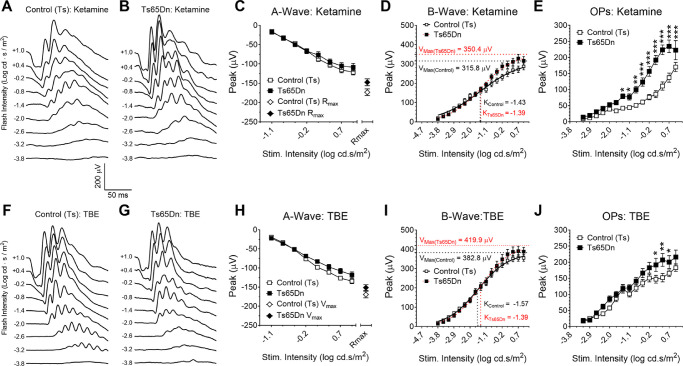
Total retinal thickness is significantly increased in Ts65Dn and Dp(16)1Yey/+ compared with control mice, because of increased thickness of inner retinal layers, including the inner nuclear layer (INL). Increased retinal vessel caliber was found in both chromosomally altered mice when compared with controls. ERG responses in Ts65Dn and Dp(16)1Yey/+ mice showed subtle alterations compared with controls. These, however, seemed to be unrelated to the thickness of the INL, but instead dependent on the anesthetic agent used (ketamine, tribromoethanol, or urethane).
We provide evidence of retinal alterations in Ts65Dn and Dp(16)1Yey/+ mice that are similar to those reported in persons with DS. Our ERG results are also a reminder that consideration should be given to the choice of anesthetic agents in such experiments.
眼部疾病是唐氏综合征(DS)最常见的合并症之一。因此,在研究 DS 的小鼠模型时,如果忽略视力的影响,可能会导致对依赖视觉系统完整性的评估结果产生误解。在这里,我们使用成像和视网膜电图(ERG)研究了两种重要的 DS 小鼠模型:Ts65Dn 和 Dp(16)1Yey/+中的眼睛结构和功能。
用裂隙灯检查角膜和前段。通过光学相干断层扫描(OCT)定量视网膜层的厚度。通过磁共振成像(MRI)测量眼睛和晶状体的尺寸。通过明场和荧光成像以及视网膜平面铺片评估视网膜血管参数。使用全视野 ERG 响应闪光刺激来评估成年小鼠的视网膜功能。
与对照小鼠相比,Ts65Dn 和 Dp(16)1Yey/+的总视网膜厚度显着增加,这是由于内视网膜层(包括内核层(INL))的厚度增加。与对照相比,在两种染色体改变的小鼠中均发现视网膜血管口径增加。与对照相比,Ts65Dn 和 Dp(16)1Yey/+小鼠的 ERG 反应显示出细微的变化。然而,这些似乎与 INL 的厚度无关,而是取决于所使用的麻醉剂(氯胺酮、三溴乙醇或尿烷)。
我们提供了 Ts65Dn 和 Dp(16)1Yey/+小鼠的视网膜改变的证据,这些改变与 DS 患者中报告的改变相似。我们的 ERG 结果也提醒人们,在这种实验中应考虑选择麻醉剂。