Huang Youhua, Zhang Ya, Liu Zetian, Liu Chuanhe, Zheng Jiaying, Qin Qiwei, Huang Xiaohong
College of Marine Sciences, South China Agricultural University, Guangzhou, China.
Guangdong Laboratory for Lingnan Modern Agriculture, Guangzhou, China.
Front Microbiol. 2020 Apr 30;11:790. doi: 10.3389/fmicb.2020.00790. eCollection 2020.
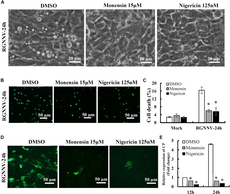
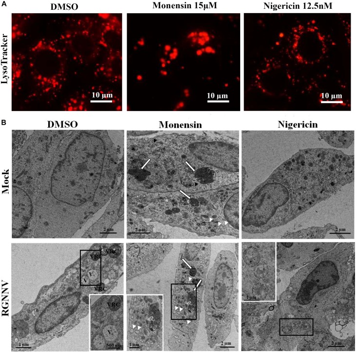
Nervous necrosis virus (NNV) is the etiological agent of viral nervous necrosis (VNN), also known as viral encephalopathy and retinopathy (VER), which results in heavy economic losses to the aquaculture industry worldwide. Dramatic cytoplasmic vacuoles were observed during NNV infection both and ; however, the origin and mechanism of cytoplasmic vacuolization remains unknown. In this report, we found that the cytoplasmic vacuole morphology became fused and enlarged during infection with red spotted grouper nervous necrosis virus (RGNNV), which was accompanied by increased cell death. Notably, Lyso-Tracker, but not Mito-Tracker or ER-Tracker, was accumulated in the vacuoles, and abnormal lysosome swelling was observed in RGNNV-infected cells, suggesting that the cytoplasmic vacuoles originated from lysosomal organelles. Cytoplasmic vacuolization and cell death in RGNNV-infected cells was completely blocked by the vacuolar H-ATPase inhibitor (bafilomycin A1), and was significantly weakened by chloroquine (CQ), a lysosomotropic agent that induces the acidification of the lysosomes. This suggests that lysosome acidification was essential for vacuole formation. Significant inhibitory effects on vacuolization and cell death were also observed in the RGNNV-infected cells following treatment with nigericin and monensin (ionophores that uncouple the proton gradient present in lysosomes). This indicated that lysosome function was tightly associated with RGNNV infection-induced cell death. In addition, vacuoles were found to be partially co-localized with GFP-LC3II punctate dots during RGNNV infection. Moreover, the severity of vacuolization and cell death were both significantly decreased after treatment with the autophagy inhibitor, 3-MA, suggesting that autophagy was involved in lysosomal vacuolization and cell death evoked by RGNNV infection. Thus, our results demonstrate that autophagy participates in lysosomal vacuolation-mediated cell death during RGNNV infection, and provides new insight into our understanding of the potential mechanisms underlying nodavirus pathogenesis .
神经坏死病毒(NNV)是病毒性神经坏死(VNN)的病原体,VNN也被称为病毒性脑病和视网膜病(VER),给全球水产养殖业造成了巨大经济损失。在NNV感染期间,均观察到显著的细胞质空泡;然而,细胞质空泡化的起源和机制仍不清楚。在本报告中,我们发现,感染红斑石斑鱼神经坏死病毒(RGNNV)期间,细胞质空泡形态融合并增大,同时细胞死亡增加。值得注意的是,溶酶体追踪染料(Lyso-Tracker)而非线粒体追踪染料(Mito-Tracker)或内质网追踪染料(ER-Tracker)在空泡中积累,并且在RGNNV感染的细胞中观察到溶酶体异常肿胀,这表明细胞质空泡起源于溶酶体细胞器。RGNNV感染细胞中的细胞质空泡化和细胞死亡被液泡H⁺-ATP酶抑制剂(巴弗洛霉素A1)完全阻断,并且被氯喹(CQ)显著减弱,氯喹是一种诱导溶酶体酸化的溶酶体促渗剂。这表明溶酶体酸化对于空泡形成至关重要。在用尼日利亚菌素和莫能菌素(使溶酶体中存在的质子梯度解偶联的离子载体)处理后的RGNNV感染细胞中,也观察到对空泡化和细胞死亡的显著抑制作用。这表明溶酶体功能与RGNNV感染诱导的细胞死亡密切相关。此外,在RGNNV感染期间,发现空泡与绿色荧光蛋白-微管相关蛋白轻链3-II(GFP-LC3II)点状结构部分共定位。此外,在用自噬抑制剂3-甲基腺嘌呤(3-MA)处理后,空泡化和细胞死亡的严重程度均显著降低,这表明自噬参与了RGNNV感染引起的溶酶体空泡化和细胞死亡。因此,我们的结果表明,自噬参与了RGNNV感染期间溶酶体空泡化介导的细胞死亡,并为我们理解诺达病毒发病机制的潜在机制提供了新的见解。