Penn Statistics in Imaging and Visualization Center, Department of Biostatistics, Epidemiology, and Informatics, Perelman School of Medicine, University of Pennsylvania, Philadelphia, PA 19104, United States.
Department of Biostatistics, Bloomberg School of Public Health, Johns Hopkins University, Baltimore, MD 21287, United States.
Neuroimage Clin. 2020;27:102256. doi: 10.1016/j.nicl.2020.102256. Epub 2020 May 16.
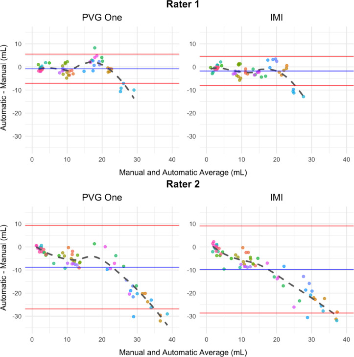
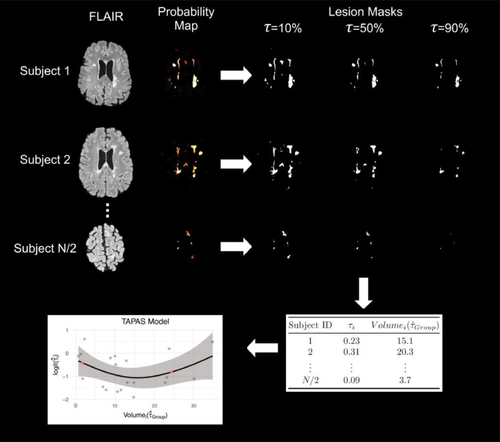
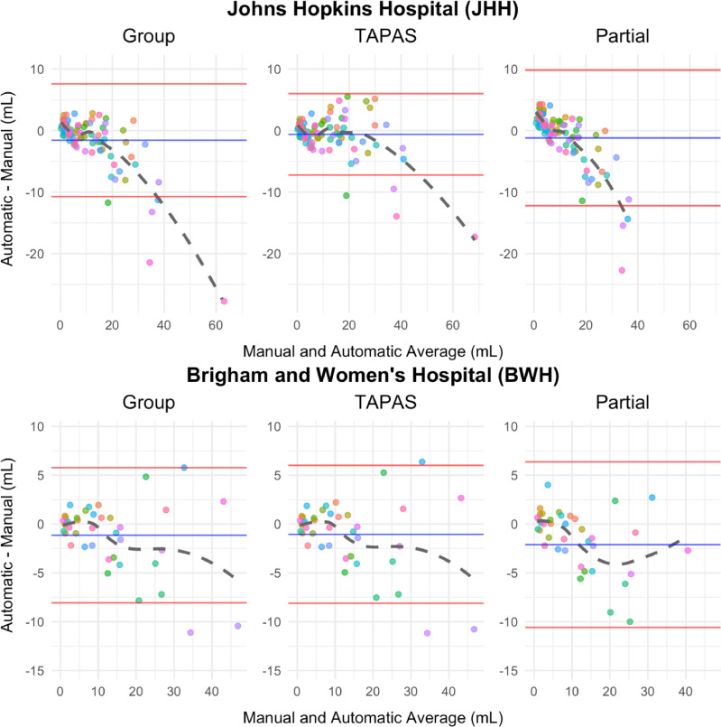
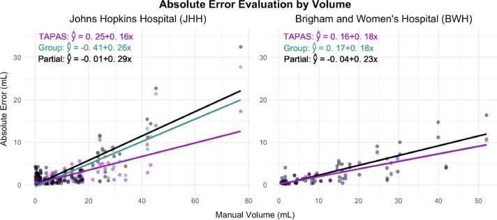
Total brain white matter lesion (WML) volume is the most widely established magnetic resonance imaging (MRI) outcome measure in studies of multiple sclerosis (MS). To estimate WML volume, there are a number of automatic segmentation methods available, yet manual delineation remains the gold standard approach. Automatic approaches often yield a probability map to which a threshold is applied to create lesion segmentation masks. Unfortunately, few approaches systematically determine the threshold employed; many methods use a manually selected threshold, thus introducing human error and bias into the automated procedure. In this study, we propose and validate an automatic thresholding algorithm, Thresholding Approach for Probability Map Automatic Segmentation in Multiple Sclerosis (TAPAS), to obtain subject-specific threshold estimates for probability map automatic segmentation of T2-weighted (T2) hyperintense WMLs. Using multimodal MRI, the proposed method applies an automatic segmentation algorithm to obtain probability maps. We obtain the true subject-specific threshold that maximizes the Sørensen-Dice similarity coefficient (DSC). Then the subject-specific thresholds are modeled on a naive estimate of volume using a generalized additive model. Applying this model, we predict a subject-specific threshold in data not used for training. We ran a Monte Carlo-resampled split-sample cross-validation (100 validation sets) using two data sets: the first obtained from the Johns Hopkins Hospital (JHH) on a Philips 3 Tesla (3T) scanner (n = 94) and a second collected at the Brigham and Women's Hospital (BWH) using a Siemens 3T scanner (n = 40). By means of the proposed automated technique, in the JHH data we found an average reduction in subject-level absolute error of 0.1 mL per one mL increase in manual volume. Using Bland-Altman analysis, we found that volumetric bias associated with group-level thresholding was mitigated when applying TAPAS. The BWH data showed similar absolute error estimates using group-level thresholding or TAPAS likely since Bland-Altman analyses indicated no systematic biases associated with group or TAPAS volume estimates. The current study presents the first validated fully automated method for subject-specific threshold prediction to segment brain lesions.
脑白质总病变体积(WML)是多发性硬化症(MS)研究中最广泛应用的磁共振成像(MRI)结果测量指标。为了估计 WML 体积,有许多自动分割方法可用,但手动勾画仍然是金标准方法。自动方法通常会生成概率图,然后对其应用阈值以创建病变分割掩模。不幸的是,很少有方法系统地确定所使用的阈值;许多方法使用手动选择的阈值,因此将人为误差和偏差引入到自动过程中。在这项研究中,我们提出并验证了一种自动阈值算法,即多发性硬化症概率图自动分割的阈值方法(TAPAS),以获得 T2 加权(T2)高信号 WML 概率图自动分割的个体特异性阈值估计。该方法使用多模态 MRI 应用自动分割算法获得概率图。我们获得了使 Sørensen-Dice 相似系数(DSC)最大化的真实个体特异性阈值。然后,使用广义加性模型对基于体积的个体特异性阈值进行建模。应用该模型,我们在未用于训练的数据中预测个体特异性阈值。我们在两个数据集上运行了蒙特卡罗重采样的分割样本交叉验证(100 个验证集):第一个数据集来自约翰霍普金斯医院(JHH)在飞利浦 3 特斯拉(3T)扫描仪上(n=94),第二个数据集在布里格姆妇女医院(BWH)使用西门子 3T 扫描仪收集(n=40)。通过所提出的自动技术,在 JHH 数据中,我们发现手动体积每增加 1 毫升,个体水平绝对误差平均减少 0.1 毫升。通过 Bland-Altman 分析,我们发现当应用 TAPAS 时,与组水平阈值相关的体积偏差得到了缓解。BWH 数据使用组水平阈值或 TAPAS 显示出相似的绝对误差估计值,可能是因为 Bland-Altman 分析表明与组或 TAPAS 体积估计值无关的系统偏差。目前的研究提出了第一个用于分割脑病变的个体特异性阈值预测的全自动化方法。