Yang Kebing, Yang Qingyan, Niu Yajuan, Fan Fengmei, Chen Song, Luo Xingguang, Tan Shuping, Wang Zhiren, Tong Jinghui, Yang Fude, Le Thang M, Li Chiang-Shan R, Tan Yunlong
Peking University Huilongguan Clinical Medical School, Beijing Huilongguan Hospital, Beijing, China.
Department of Psychiatry, Yale University School of Medicine, New Haven, CT, United States.
Front Psychiatry. 2020 May 5;11:364. doi: 10.3389/fpsyt.2020.00364. eCollection 2020.
Many studies reported structural brain changes in patients with alcohol dependence (PADs). However, no studies identified structural correlates of apathy that might aggravate alcohol misuse. Here, we explored regional differences in cortical thickness in PADs relative to healthy controls (HCs), and examined the potential correlation of regional thickness with the severity of apathy.
Magnetic resonance imaging data were collected from 33 male PADs and 35 male age- and education-matched HCs. We used the FreeSurfer software to investigate group differences in cortical thickness across 148 regions. Apathy was evaluated using the Lille Apathy Rating Scale-Informant (LARS-I). Regression analyses examined the relationship between cortical thickness of regions of interest and apathy score in PADs.
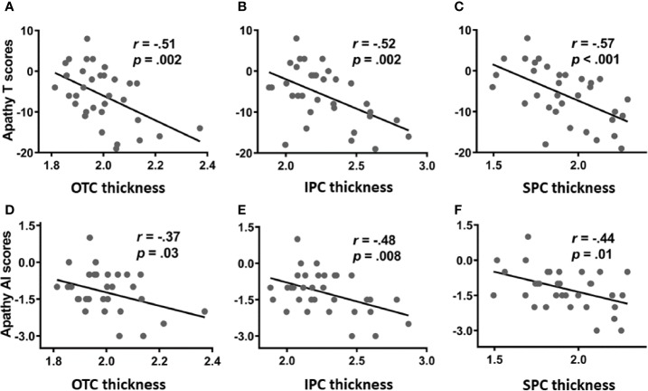
Compared to HCs, PADs showed significant decreases in the cortical thickness of occipito-temporal cortex (OTC), including the left middle occipital gyrus and occipital pole, right superior occipital gyri, and bilateral lingual gyrus; bilateral superior parietal cortex (SPC), including the right intraparietal sulcus; and bilateral inferior parietal cortex (IPC). Furthermore, the cortical thickness of all of the three regions was negatively correlated with the apathy total scores. The cortical thickness of the IPC was also negatively correlated with the action initiation subscore of the LARS-I.
The current results suggest the thickness of bilateral parietal and occipital temporal cortices as neural markers of apathy in PADs. These findings add to the literature by identifying the neural bases of a critical clinical feature of individuals with alcoholism.
许多研究报道了酒精依赖患者(PADs)的脑结构变化。然而,尚无研究确定可能加重酒精滥用的冷漠情绪的结构相关性。在此,我们探讨了PADs相对于健康对照者(HCs)在皮质厚度上的区域差异,并研究了区域厚度与冷漠严重程度之间的潜在相关性。
收集了33名男性PADs和35名年龄及教育程度匹配的男性HCs的磁共振成像数据。我们使用FreeSurfer软件研究148个区域的皮质厚度的组间差异。使用里尔冷漠评定量表- informant版(LARS-I)评估冷漠情绪。回归分析检验了PADs中感兴趣区域的皮质厚度与冷漠评分之间的关系。
与HCs相比,PADs在枕颞叶皮质(OTC)的皮质厚度显著降低,包括左侧枕中回和枕极、右侧枕上回以及双侧舌回;双侧顶上叶皮质(SPC),包括右侧顶内沟;以及双侧顶下叶皮质(IPC)。此外,这三个区域的皮质厚度均与冷漠总分呈负相关。IPC的皮质厚度也与LARS-I的行动启动子评分呈负相关。
目前的结果表明双侧顶叶和枕颞叶皮质的厚度是PADs中冷漠情绪的神经标志物。这些发现通过确定酒精中毒个体关键临床特征的神经基础,为该领域的文献增添了内容。