Lopa Silvia, Piraino Francesco, Talò Giuseppe, Mainardi Valerio Luca, Bersini Simone, Pierro Margherita, Zagra Luigi, Rasponi Marco, Moretti Matteo
IRCCS Istituto Ortopedico Galeazzi, Cell and Tissue Engineering Laboratory, Milan, Italy.
Department of Electronics, Information and Bioengineering, Politecnico di Milano, Milan, Italy.
Front Bioeng Biotechnol. 2020 May 5;8:366. doi: 10.3389/fbioe.2020.00366. eCollection 2020.
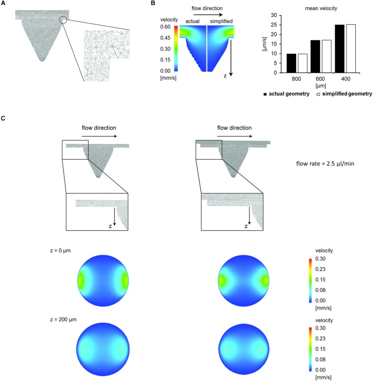
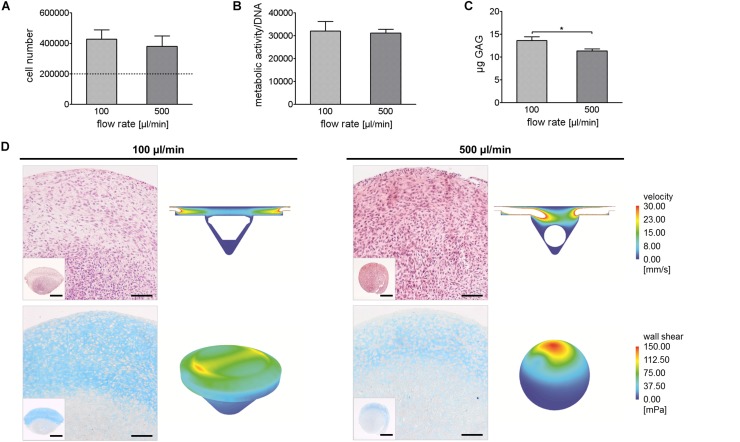
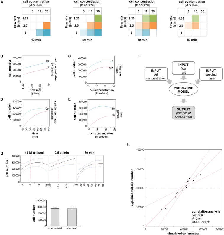
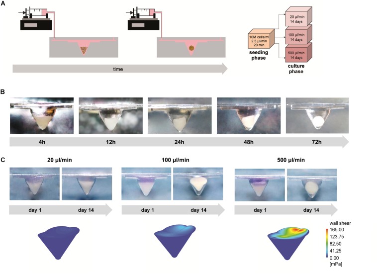
Three-dimensional (3D) cell spheroids are being increasingly applied in many research fields due to their enhanced biological functions as compared to conventional two-dimensional (2D) cultures. 3D cell spheroids can replicate tissue functions, which enables their use both as models and as building blocks in tissue biofabrication approaches. In this study, we developed a perfusable microfluidic platform suitable for robust and reproducible 3D cell spheroid formation and tissue maturation. The geometry of the device was optimized through computational fluid dynamic (CFD) simulations to improve cell trapping. Experimental data were used in turn to generate a model able to predict the number of trapped cells as a function of cell concentration, flow rate, and seeding time. We demonstrated that tuning non-geometrical parameters it is possible to control the size and shape of 3D cell spheroids generated using articular chondrocytes (ACs) as cellular model. After seeding, cells were cultured under perfusion at different flow rates (20, 100, and 500 μl/min), which induced the formation of conical and spherical spheroids. Wall shear stress values on cell spheroids, computed by CFD simulations, increased accordingly to the flow rate while remaining under the chondroprotective threshold in all configurations. The effect of flow rate on cell number, metabolic activity, and tissue-specific matrix deposition was evaluated and correlated with fluid velocity and shear stress distribution. The obtained results demonstrated that our device represents a helpful tool to generate stable 3D cell spheroids which can find application both to develop advanced models for the study of physio-pathological tissue maturation mechanisms and to obtain building blocks for the biofabrication of macrotissues.
与传统的二维(2D)培养相比,三维(3D)细胞球体因其增强的生物学功能而在许多研究领域中得到越来越广泛的应用。3D细胞球体可以复制组织功能,这使其既可以用作模型,也可以用作组织生物制造方法中的构建块。在本研究中,我们开发了一种可灌注的微流控平台,适用于稳健且可重复的3D细胞球体形成和组织成熟。通过计算流体动力学(CFD)模拟对设备的几何形状进行了优化,以改善细胞捕获。反过来,实验数据被用于生成一个模型,该模型能够预测捕获细胞的数量作为细胞浓度、流速和接种时间的函数。我们证明,通过调整非几何参数,可以控制以关节软骨细胞(ACs)为细胞模型生成的3D细胞球体的大小和形状。接种后,细胞在不同流速(20、100和500 μl/min)下进行灌注培养,这诱导了锥形和球形球体的形成。通过CFD模拟计算得到的细胞球体上的壁面剪应力值随流速相应增加,同时在所有配置下均保持在软骨保护阈值以下。评估了流速对细胞数量、代谢活性和组织特异性基质沉积的影响,并将其与流体速度和剪应力分布相关联。所得结果表明,我们的设备是生成稳定3D细胞球体的有用工具,这些球体既可以用于开发用于研究生理病理组织成熟机制的先进模型,也可以用于获得宏观组织生物制造的构建块。