Yu Nan, Yang Chuangbo, Ma Guangming, Dang Shan, Ren Zhanli, Wang Shaoyu, Yu Yong
Department of Radiology, The affiliated hospital of Chinese traditional medical university, Xian Yang China, -2# Weiyang Western Road, Xian Yang, 712000, China.
Department of Medical Technology, The affiliated hospital of Chinese traditional medical university, Xian Yang, China.
BMC Med Imaging. 2020 May 20;20(1):53. doi: 10.1186/s12880-020-00451-w.
To assess the feasibility of various magnetic resonance imaging (MRI) sequences for the detection of pulmonary nodules by comparing the detection rate of computed tomography (CT).
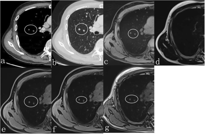
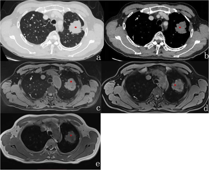
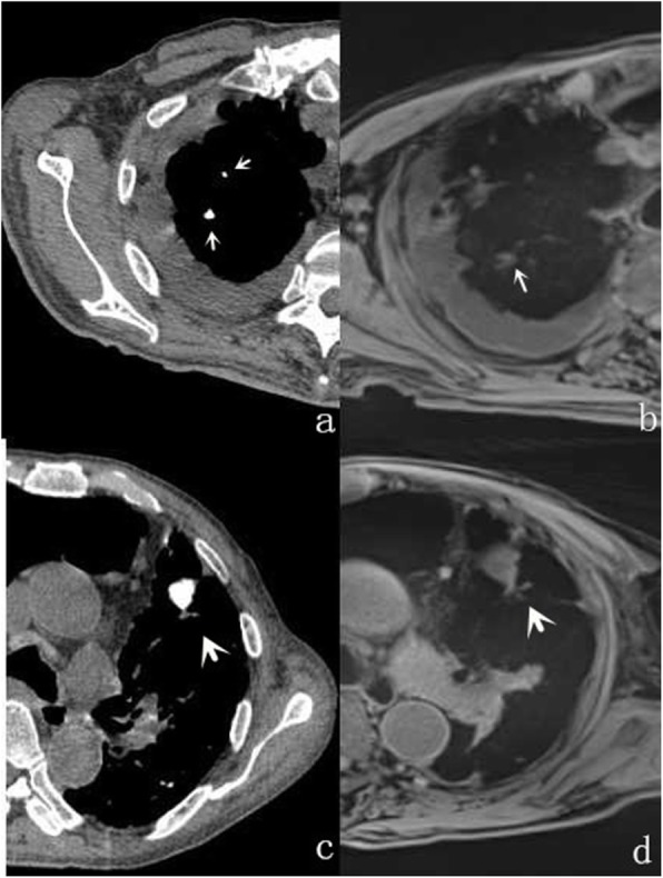
Forty-two patients with pulmonary nodules detected by multi-slice CT (MSCT) were prospectively enrolled in the present study between November 2016 and February 2017. Chest MRI was acquired within 24 h of CT. The MRI protocol included free-breathing radial VIBE (r-VIBE) and a conventional breathhold T1-weighted VIBE (C-VIBE) were analyzed by two independent radiologists. Both detection and morphology results of each MRI image were recorded. Subjective image evaluation in terms of overall nodule morphology on the MRI images was carried out using the 4-point scoring criteria. The MRI results were compared with those from CT, with the results of MSCT serving as the reference standard.
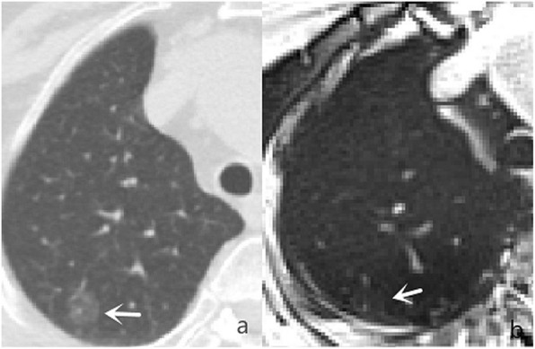
Two hundred and fifty-eight solid pulmonary nodules in 42 patients were detected by CT. The r-VIBE correctly detected 94% of the pulmonary nodules as compared with CT. The detection rate increased to 100% for lesions ≥6 mm. The C-VIBE had a lower overall detection rate (64.3%) of pulmonary nodules. The difference in the subjective image evaluation scores between the two sequences was statistically significant (p < 0.001).
Significantly increased detection rates were obtained with free-breathing r-VIBE as compared with C-VIBE for the detection of pulmonary nodules and also provided more information when evaluating the nodules as compared with C-VIBE.
通过比较计算机断层扫描(CT)的检测率,评估各种磁共振成像(MRI)序列检测肺结节的可行性。
2016年11月至2017年2月期间,前瞻性纳入42例经多层CT(MSCT)检测出肺结节的患者。在CT检查后24小时内进行胸部MRI检查。MRI检查方案包括自由呼吸径向容积内插屏气检查法(r-VIBE)和传统屏气T1加权容积内插屏气检查法(C-VIBE),由两名独立的放射科医生进行分析。记录每个MRI图像的检测结果和形态学结果。使用4分评分标准对MRI图像上的整体结节形态进行主观图像评估。将MRI结果与CT结果进行比较,以MSCT结果作为参考标准。
CT检测出42例患者中的258个实性肺结节。与CT相比,r-VIBE正确检测出94%的肺结节。对于≥6mm的病变,检测率提高到100%。C-VIBE对肺结节的总体检测率较低(64.3%)。两个序列之间主观图像评估得分的差异具有统计学意义(p<0.001)。
与C-VIBE相比,自由呼吸r-VIBE在检测肺结节时检测率显著提高,并且在评估结节时比C-VIBE提供了更多信息。