Parikh Aaroh M, Grogan Raymon H, Morón Fanny E
School of Medicine, Baylor College of Medicine, Houston, TX 77030, USA.
Department of Internal Medicine, Santa Clara Valley Medical Center, San Jose, CA 95128, USA.
Int J Endocrinol. 2020 Jan 25;2020:9649564. doi: 10.1155/2020/9649564. eCollection 2020.
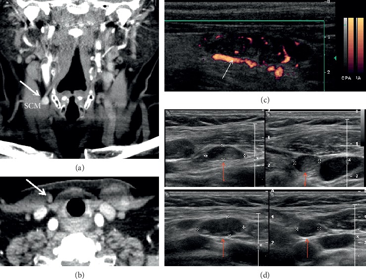
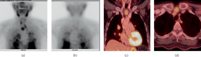
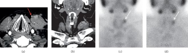
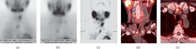
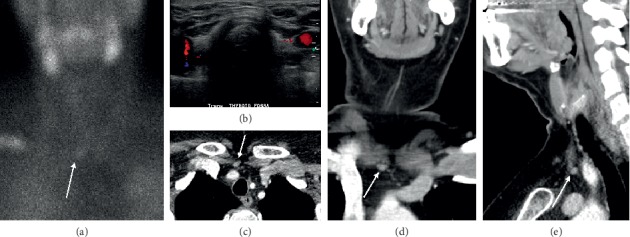
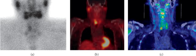
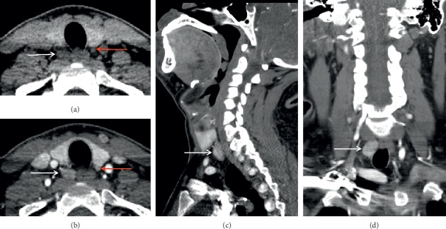
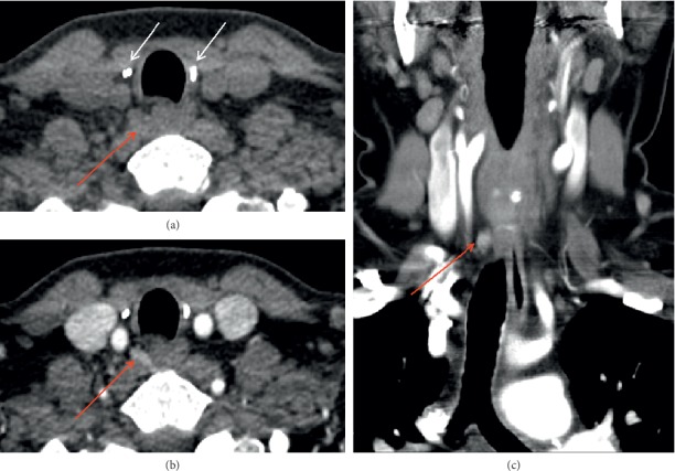
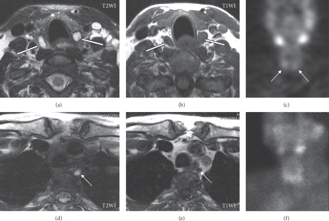
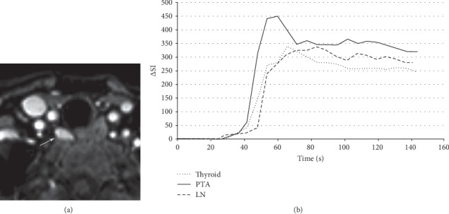
The localization of persistent or recurrent disease in reoperative patients with primary hyperparathyroidism presents challenges for radiologists and surgeons alike. In this article, we summarize the relevant imaging modalities, compare their accuracy in identifying reoperative disease, and outline their advantages and disadvantages. Accurate localization by preoperative imaging is a predictor of operative success, whereas negative or discordant preoperative imaging is a risk factor for operative failure. Ultrasound is a common first-line modality because it is inexpensive, accessible, and radiation-free. However, it is highly operator-dependent and less accurate in the reoperative setting than in the primary setting. Sestamibi scintigraphy is superior to ultrasound in localizing reoperative disease but requires radiation, prolonged imaging times, and reader experience for accurate interpretation. Like ultrasound, sestamibi scintigraphy is less accurate in the reoperative setting because reoperative patients can exhibit distorted anatomy, altered perfusion of remaining glands, and interference of radiotracer uptake. Meanwhile, four-dimensional computed tomography (4DCT) is superior to ultrasound and sestamibi scintigraphy in localizing reoperative disease but requires the use of radiation and intravenous contrast. Both 4DCT and magnetic resonance imaging (MRI) do not significantly differ in accuracy between unexplored and reoperative patients. However, MRI is more costly, inaccessible, and time-consuming than 4DCT and is inappropriate as a first-line modality. Hybrid imaging with positron emission tomography and computed tomography (PET/CT) may be a promising second-line modality in the reoperative setting, particularly when first-line modalities are discordant or inconclusive. Lastly, selective venous sampling should be reserved for challenging cases in which noninvasive modalities are negative or discordant. In the challenging population of reoperative patients with PHPT, a multimodality approach that utilizes the expertise of high-volume centers can accurately localize persistent or recurrent disease and enable curative parathyroidectomy.
对于再次手术的原发性甲状旁腺功能亢进患者,持续性或复发性疾病的定位给放射科医生和外科医生都带来了挑战。在本文中,我们总结了相关的成像方式,比较了它们在识别再次手术疾病方面的准确性,并概述了它们的优缺点。术前成像的准确定位是手术成功的预测指标,而术前成像结果为阴性或不一致则是手术失败的风险因素。超声是一种常见的一线检查方式,因为它价格低廉、易于获得且无辐射。然而,它高度依赖操作人员,在再次手术情况下的准确性低于初次手术情况。锝[99mTc]甲氧基异丁基异腈闪烁扫描在定位再次手术疾病方面优于超声,但需要辐射、较长的成像时间,且准确解读需要阅片者的经验。与超声一样,锝[99mTc]甲氧基异丁基异腈闪烁扫描在再次手术情况下的准确性较低,因为再次手术患者可能存在解剖结构变形、剩余腺体灌注改变以及放射性示踪剂摄取的干扰。同时,四维计算机断层扫描(4DCT)在定位再次手术疾病方面优于超声和锝[99mTc]甲氧基异丁基异腈闪烁扫描,但需要使用辐射和静脉造影剂。4DCT和磁共振成像(MRI)在未进行过手术和再次手术患者中的准确性没有显著差异。然而,MRI比4DCT成本更高、不易获得且耗时更长,不适合作为一线检查方式。正电子发射断层扫描和计算机断层扫描(PET/CT)的联合成像在再次手术情况下可能是一种有前景的二线检查方式,特别是当一线检查方式结果不一致或不确定时。最后,选择性静脉采血应保留用于无创检查方式结果为阴性或不一致的疑难病例。在具有挑战性的再次手术的原发性甲状旁腺功能亢进患者群体中,采用多模式方法并利用大容量中心的专业知识,可以准确地定位持续性或复发性疾病,并实现治愈性甲状旁腺切除术。