Department of Internal Medicine Medical University of South Carolina Charleston SC.
Ralph H. Johnson Veterans Affairs Medical Center Charleston SC.
J Am Heart Assoc. 2020 Jun 16;9(12):e014542. doi: 10.1161/JAHA.119.014542. Epub 2020 Jun 1.
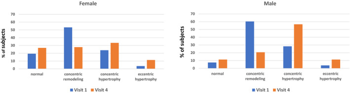
Background Progressive cardiac remodeling and worsening myocardial function over time have been proposed as potential mediators of heart failure in obesity. Methods and Results We serially assessed cardiac structure and function in 254 subjects participating in a longitudinal study of obesity. Demographic, clinical, laboratory, and echocardiographic features were determined at baseline and 2-, 6-, and 11-year follow-up. We measured body mass index (BMI) exposure as the area under the curve of the BMI at each of the 4 visits. At enrollment, mean age of the subjects was 47 years, 79% were women, mean BMI was 44 kg/m, 26% had diabetes mellitus, 48% had hypertension, and 53% had hyperlipidemia. Between baseline and 11 years, BMI increased by 1.1 and 0.3 kg/m in men and women, respectively. There were modest increases in left ventricular (LV) end-diastolic volume, LV mass, and left atrial volume, and significant decreases in early/late mitral diastolic flow velocity ratio and E wave deceleration time. However, there were no significant changes in LV ejection fraction or ratio of early mitral diastolic flow velocity/early mitral annular velocity, whereas right ventricular fractional area change increased. Significant predictors of the change in LV mass were male sex, baseline BMI, BMI area under the curve, and change in LV stroke volume, but not smoking, hypertension, or diabetes mellitus. Conclusions In long-standing, persistent severe obesity, there was evidence of cardiac remodeling over a period of 11 years, but no clear worsening of systolic or diastolic function. Measures of remodeling were most strongly related to BMI. The observed changes might predispose to heart failure with preserved ejection fraction, but are not classic for an evolving dilated cardiomyopathy.
随着时间的推移,心脏进行性重构和心肌功能恶化被认为是肥胖导致心力衰竭的潜在介质。
我们对 254 名参与肥胖纵向研究的患者进行了连续心脏结构和功能评估。在基线、2 年、6 年和 11 年随访时,确定了人口统计学、临床、实验室和超声心动图特征。我们将体重指数(BMI)暴露定义为 4 次访视时 BMI 的曲线下面积。在入组时,患者的平均年龄为 47 岁,79%为女性,平均 BMI 为 44kg/m2,26%患有糖尿病,48%患有高血压,53%患有高脂血症。在基线到 11 年期间,男性和女性的 BMI 分别增加了 1.1kg/m2 和 0.3kg/m2。左心室(LV)舒张末期容积、LV 质量和左心房容积适度增加,早期/晚期二尖瓣舒张血流速度比和 E 波减速时间显著降低。然而,LV 射血分数或早期二尖瓣舒张血流速度/早期二尖瓣环速度的比值没有显著变化,而右心室分数区域变化增加。LV 质量变化的显著预测因素为男性、基线 BMI、BMI 曲线下面积和 LV 每搏量的变化,而不是吸烟、高血压或糖尿病。
在长期、持续的严重肥胖中,在 11 年期间有心脏重构的证据,但没有明显的收缩或舒张功能恶化。重构的测量与 BMI 关系最密切。观察到的变化可能导致射血分数保留的心力衰竭,但不是进展性扩张型心肌病的典型特征。