• 文献检索
  • 文档翻译
  • 深度研究
  • 学术资讯
  • Suppr Zotero 插件Zotero 插件
  • 邀请有礼
  • 套餐&价格
  • 历史记录
应用&插件
Suppr Zotero 插件Zotero 插件浏览器插件Mac 客户端Windows 客户端微信小程序
定价
高级版会员购买积分包购买API积分包
服务
文献检索文档翻译深度研究API 文档MCP 服务
关于我们
关于 Suppr公司介绍联系我们用户协议隐私条款
关注我们

Suppr 超能文献

核心技术专利:CN118964589B侵权必究
粤ICP备2023148730 号-1Suppr @ 2026

文献检索

告别复杂PubMed语法,用中文像聊天一样搜索,搜遍4000万医学文献。AI智能推荐,让科研检索更轻松。

立即免费搜索

文件翻译

保留排版,准确专业,支持PDF/Word/PPT等文件格式,支持 12+语言互译。

免费翻译文档

深度研究

AI帮你快速写综述,25分钟生成高质量综述,智能提取关键信息,辅助科研写作。

立即免费体验

使用22层U型网络对数字化头颈部组织学图像上的鳞状细胞癌进行分割。

Using a 22-Layer U-Net to Perform Segmentation of Squamous Cell Carcinoma on Digitized Head and Neck Histological Images.

作者信息

Mavuduru Amol, Halicek Martin, Shahedi Maysam, Little James V, Chen Amy Y, Myers Larry L, Fei Baowei

机构信息

Dept. of Bioengineering, University of Texas at Dallas, TX.

Dept. of Computer Science, University of Texas at Dallas, TX.

出版信息

Proc SPIE Int Soc Opt Eng. 2020 Feb;11320. doi: 10.1117/12.2549061. Epub 2020 Mar 16.

DOI:10.1117/12.2549061
PMID:32476709
原文链接:https://pmc.ncbi.nlm.nih.gov/articles/PMC7261613/
Abstract

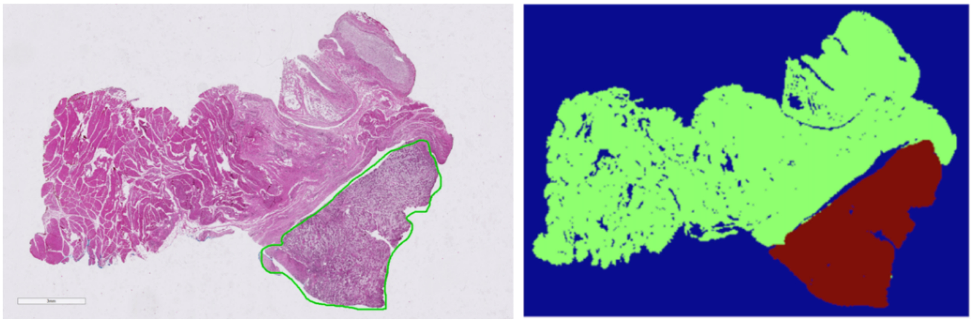
Squamous cell carcinoma (SCC) comprises over 90 percent of tumors in the head and neck. The diagnosis process involves performing surgical resection of tissue and creating histological slides from the removed tissue. Pathologists detect SCC in histology slides, and may fail to correctly identify tumor regions within the slides. In this study, a dataset of patches extracted from 200 digitized histological images from 84 head and neck SCC patients was used to train, validate and test the segmentation performance of a fully-convolutional U-Net architecture. The neural network achieved a pixel-level segmentation AUC of 0.89 on the testing group. The average segmentation time for whole slide images was 72 seconds. The training, validation, and testing process in this experiment produces a model that has the potential to help segment SCC images in histological images with improved speed and accuracy compared to the manual segmentation process performed by pathologists.

摘要

鳞状细胞癌(SCC)占头颈部肿瘤的90%以上。诊断过程包括对组织进行手术切除,并从切除的组织中制作组织学切片。病理学家在组织学切片中检测SCC,可能无法正确识别切片内的肿瘤区域。在本研究中,使用从84名头颈部SCC患者的200张数字化组织学图像中提取的图像块数据集来训练、验证和测试全卷积U-Net架构的分割性能。该神经网络在测试组上实现了0.89的像素级分割AUC。全幻灯片图像的平均分割时间为72秒。与病理学家进行的手动分割过程相比,本实验中的训练、验证和测试过程产生了一个有潜力帮助在组织学图像中分割SCC图像的模型,其速度和准确性更高。

https://cdn.ncbi.nlm.nih.gov/pmc/blobs/c204/7261613/837a1f69690a/nihms-1592297-f0004.jpg
https://cdn.ncbi.nlm.nih.gov/pmc/blobs/c204/7261613/b402e7b608c5/nihms-1592297-f0001.jpg
https://cdn.ncbi.nlm.nih.gov/pmc/blobs/c204/7261613/ea98fb7db696/nihms-1592297-f0002.jpg
https://cdn.ncbi.nlm.nih.gov/pmc/blobs/c204/7261613/a8206efea08a/nihms-1592297-f0003.jpg
https://cdn.ncbi.nlm.nih.gov/pmc/blobs/c204/7261613/837a1f69690a/nihms-1592297-f0004.jpg

相似文献

1
Using a 22-Layer U-Net to Perform Segmentation of Squamous Cell Carcinoma on Digitized Head and Neck Histological Images.使用22层U型网络对数字化头颈部组织学图像上的鳞状细胞癌进行分割。
Proc SPIE Int Soc Opt Eng. 2020 Feb;11320. doi: 10.1117/12.2549061. Epub 2020 Mar 16.
2
Detection of Squamous Cell Carcinoma in Digitized Histological Images from the Head and Neck Using Convolutional Neural Networks.使用卷积神经网络检测头颈部数字化组织学图像中的鳞状细胞癌。
Proc SPIE Int Soc Opt Eng. 2019 Feb;10956. doi: 10.1117/12.2512570. Epub 2019 Mar 18.
3
Head and Neck Cancer Detection in Digitized Whole-Slide Histology Using Convolutional Neural Networks.基于卷积神经网络的数字化全切片组织学中的头颈部癌症检测
Sci Rep. 2019 Oct 1;9(1):14043. doi: 10.1038/s41598-019-50313-x.
4
A machine learning model for separating epithelial and stromal regions in oral cavity squamous cell carcinomas using H&E-stained histology images: A multi-center, retrospective study.基于 H&E 染色组织学图像的机器学习模型在口腔鳞状细胞癌上皮和间质区域分离中的应用:一项多中心回顾性研究。
Oral Oncol. 2022 Aug;131:105942. doi: 10.1016/j.oraloncology.2022.105942. Epub 2022 Jun 8.
5
AnatomyNet: Deep learning for fast and fully automated whole-volume segmentation of head and neck anatomy.AnatomyNet:用于快速和全自动对头颈部解剖结构进行整体体积分割的深度学习方法。
Med Phys. 2019 Feb;46(2):576-589. doi: 10.1002/mp.13300. Epub 2018 Dec 17.
6
Influence of Data Augmentation Strategies on the Segmentation of Oral Histological Images Using Fully Convolutional Neural Networks.基于全卷积神经网络的数据增强策略对口腔组织学图像分割的影响。
J Digit Imaging. 2023 Aug;36(4):1608-1623. doi: 10.1007/s10278-023-00814-z. Epub 2023 Apr 3.
7
Oral squamous cell carcinoma diagnosis in digitized histological images using convolutional neural network.利用卷积神经网络对数字化组织学图像进行口腔鳞状细胞癌诊断。
J Dent Sci. 2023 Jan;18(1):322-329. doi: 10.1016/j.jds.2022.08.017. Epub 2022 Sep 8.
8
Convolutional neural network for automated mass segmentation in mammography.卷积神经网络在乳腺 X 线摄影中用于自动肿块分割。
BMC Bioinformatics. 2020 Dec 9;21(Suppl 1):192. doi: 10.1186/s12859-020-3521-y.
9
A Fully Automated Artificial Intelligence System to Assist Pathologists' Diagnosis to Predict Histologically High-grade Urothelial Carcinoma from Digitized Urine Cytology Slides Using Deep Learning.一种全自动人工智能系统,利用深度学习从数字化尿液细胞学载玻片辅助病理学家诊断预测组织学高级别尿路上皮癌。
Eur Urol Oncol. 2024 Apr;7(2):258-265. doi: 10.1016/j.euo.2023.11.009. Epub 2023 Dec 7.
10
Cyto R-CNN and CytoNuke Dataset: Towards reliable whole-cell segmentation in bright-field histological images.细胞 R-CNN 和细胞核分割数据集:实现亮场组织学图像中可靠的全细胞分割。
Comput Methods Programs Biomed. 2024 Jul;252:108215. doi: 10.1016/j.cmpb.2024.108215. Epub 2024 May 11.

引用本文的文献

1
Artificial Intelligence in Head and Neck Surgery.人工智能在头颈部外科中的应用。
Otolaryngol Clin North Am. 2024 Oct;57(5):803-820. doi: 10.1016/j.otc.2024.05.001. Epub 2024 Jun 22.
2
Artificial intelligence in head and neck cancer diagnosis.人工智能在头颈癌诊断中的应用
J Pathol Inform. 2022 Nov 8;13:100153. doi: 10.1016/j.jpi.2022.100153. eCollection 2022.
3
Hyperspectral Microscopic Imaging for the Detection of Head and Neck Squamous Cell Carcinoma on Histologic Slides.用于在组织学切片上检测头颈部鳞状细胞癌的高光谱显微成像

本文引用的文献

1
Head and Neck Cancer Detection in Digitized Whole-Slide Histology Using Convolutional Neural Networks.基于卷积神经网络的数字化全切片组织学中的头颈部癌症检测
Sci Rep. 2019 Oct 1;9(1):14043. doi: 10.1038/s41598-019-50313-x.
2
Tumor margin assessment of surgical tissue specimen of cancer patients using label-free hyperspectral imaging.使用无标记高光谱成像技术对癌症患者手术组织标本进行肿瘤边缘评估。
Proc SPIE Int Soc Opt Eng. 2017 Jan-Feb;10054. doi: 10.1117/12.2249803. Epub 2017 Feb 14.
3
Label-free reflectance hyperspectral imaging for tumor margin assessment: a pilot study on surgical specimens of cancer patients.
Proc SPIE Int Soc Opt Eng. 2021 Feb;11603. doi: 10.1117/12.2581970. Epub 2021 Feb 15.
4
Artificial Intelligence-Based Segmentation of Residual Tumor in Histopathology of Pancreatic Cancer after Neoadjuvant Treatment.基于人工智能的新辅助治疗后胰腺癌组织病理学中残留肿瘤的分割
Cancers (Basel). 2021 Oct 12;13(20):5089. doi: 10.3390/cancers13205089.
5
Comparison of texture-based classification and deep learning for plantar soft tissue histology segmentation.基于纹理分类与深度学习的足底软组织组织病理学分割比较。
Comput Biol Med. 2021 Jul;134:104491. doi: 10.1016/j.compbiomed.2021.104491. Epub 2021 May 15.
用于肿瘤边缘评估的无标记反射率高光谱成像:癌症患者手术标本的初步研究
J Biomed Opt. 2017 Aug;22(8):1-7. doi: 10.1117/1.JBO.22.8.086009.
4
Head and Neck Squamous Cell Carcinoma: Update on Epidemiology, Diagnosis, and Treatment.头颈部鳞状细胞癌:流行病学、诊断和治疗的最新进展
Mayo Clin Proc. 2016 Mar;91(3):386-96. doi: 10.1016/j.mayocp.2015.12.017.