Janssen Boris V, Theijse Rutger, van Roessel Stijn, de Ruiter Rik, Berkel Antonie, Huiskens Joost, Busch Olivier R, Wilmink Johanna W, Kazemier Geert, Valkema Pieter, Farina Arantza, Verheij Joanne, de Boer Onno J, Besselink Marc G
Department of Surgery, Amsterdam UMC, Cancer Center Amsterdam, University of Amsterdam, 1081 HV Amsterdam, The Netherlands.
Department of Pathology, Amsterdam UMC, Cancer Center Amsterdam, University of Amsterdam, 1081 HV Amsterdam, The Netherlands.
Cancers (Basel). 2021 Oct 12;13(20):5089. doi: 10.3390/cancers13205089.
Histologic examination of resected pancreatic cancer after neoadjuvant therapy (NAT) is used to assess the effect of NAT and may guide the choice for adjuvant treatment. However, evaluating residual tumor burden in pancreatic cancer is challenging given tumor response heterogeneity and challenging histomorphology. Artificial intelligence techniques may offer a more reproducible approach.
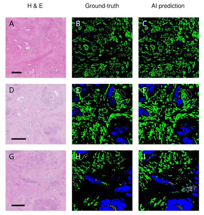
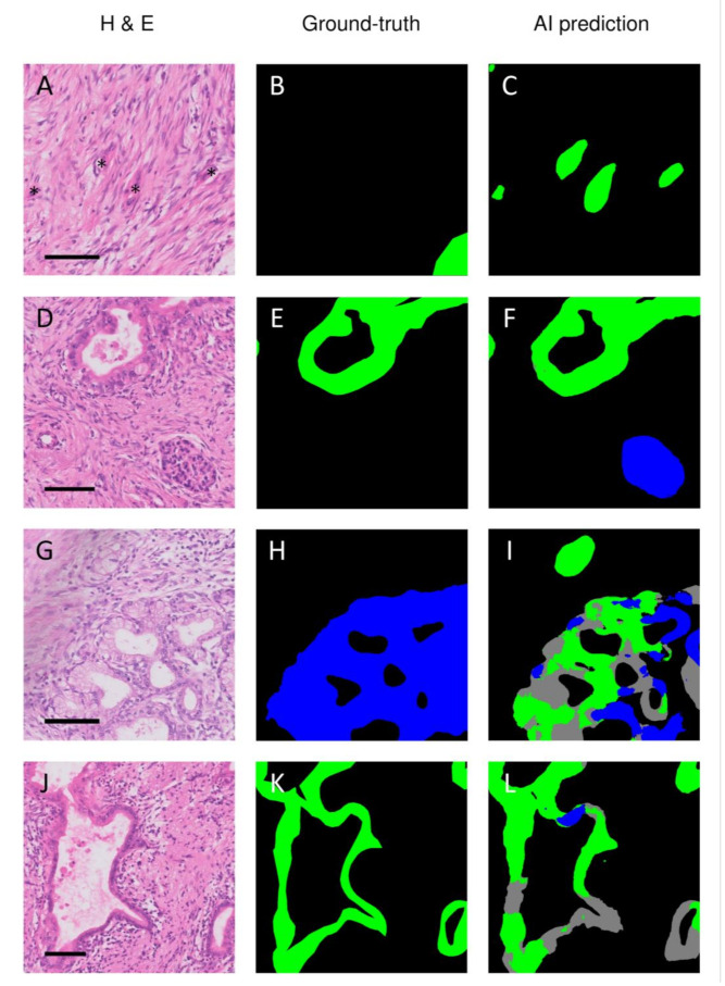
From 64 patients, one H&E-stained slide of resected pancreatic cancer after NAT was digitized. Three separate classes were manually outlined in each slide (i.e., tumor, normal ducts, and remaining epithelium). Corresponding segmentation masks and patches were generated and distributed over training, validation, and test sets. Modified U-nets with varying encoders were trained, and F1 scores were obtained to express segmentation accuracy.
The highest mean segmentation accuracy was obtained using modified U-nets with a DenseNet161 encoder. Tumor tissue was segmented with a high mean F1 score of 0.86, while the overall multiclass average F1 score was 0.82.
This study shows that artificial intelligence-based assessment of residual tumor burden is feasible given the promising obtained F1 scores for tumor segmentation. This model could be developed into a tool for the objective evaluation of the response to NAT and may potentially guide the choice for adjuvant treatment.
新辅助治疗(NAT)后切除的胰腺癌的组织学检查用于评估NAT的效果,并可能指导辅助治疗的选择。然而,鉴于肿瘤反应的异质性和具有挑战性的组织形态学,评估胰腺癌的残留肿瘤负荷具有挑战性。人工智能技术可能提供一种更具可重复性的方法。
从64例患者中,将一张NAT后切除的胰腺癌的苏木精-伊红(H&E)染色切片进行数字化处理。在每张切片中手动勾勒出三个不同的类别(即肿瘤、正常导管和剩余上皮)。生成相应的分割掩码和图像块,并分配到训练集、验证集和测试集中。使用不同编码器的改良U-Net进行训练,并获得F1分数以表示分割准确性。
使用具有DenseNet161编码器的改良U-Net获得了最高的平均分割准确性。肿瘤组织的平均F1分数较高,为0.86,而整体多类别平均F1分数为0.82。
本研究表明,鉴于在肿瘤分割中获得了有前景的F1分数,基于人工智能的残留肿瘤负荷评估是可行的。该模型可以开发成为一种客观评估NAT反应的工具,并可能潜在地指导辅助治疗的选择。