• 文献检索
  • 文档翻译
  • 深度研究
  • 学术资讯
  • Suppr Zotero 插件Zotero 插件
  • 邀请有礼
  • 套餐&价格
  • 历史记录
应用&插件
Suppr Zotero 插件Zotero 插件浏览器插件Mac 客户端Windows 客户端微信小程序
定价
高级版会员购买积分包购买API积分包
服务
文献检索文档翻译深度研究API 文档MCP 服务
关于我们
关于 Suppr公司介绍联系我们用户协议隐私条款
关注我们

Suppr 超能文献

核心技术专利:CN118964589B侵权必究
粤ICP备2023148730 号-1Suppr @ 2026

文献检索

告别复杂PubMed语法,用中文像聊天一样搜索,搜遍4000万医学文献。AI智能推荐,让科研检索更轻松。

立即免费搜索

文件翻译

保留排版,准确专业,支持PDF/Word/PPT等文件格式,支持 12+语言互译。

免费翻译文档

深度研究

AI帮你快速写综述,25分钟生成高质量综述,智能提取关键信息,辅助科研写作。

立即免费体验

CT衍生的亨氏单位:一种确定受辐照患者骨质疏松症和辐射相关骨折风险的简便方法。

CT Derived Hounsfield Unit: An Easy Way to Determine Osteoporosis and Radiation Related Fracture Risk in Irradiated Patients.

作者信息

Yaprak Gokhan, Gemici Cengiz, Seseogullari Ozgur O, Karabag Irem S, Cini Nilsu

机构信息

Department of Radiation Oncology, Kartal Dr. Lutfi Kirdar Education and Research Hospital, Istanbul, Turkey.

Department of Radiation Oncology, Biruni University Medicana Hospital, Istanbul, Turkey.

出版信息

Front Oncol. 2020 May 13;10:742. doi: 10.3389/fonc.2020.00742. eCollection 2020.

DOI:10.3389/fonc.2020.00742
PMID:32477951
原文链接:https://pmc.ncbi.nlm.nih.gov/articles/PMC7237579/
Abstract

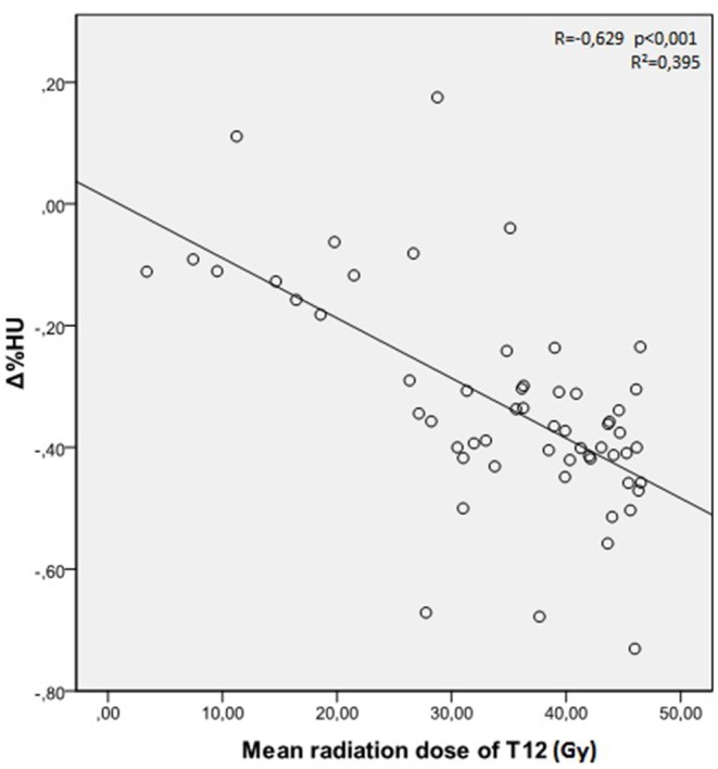
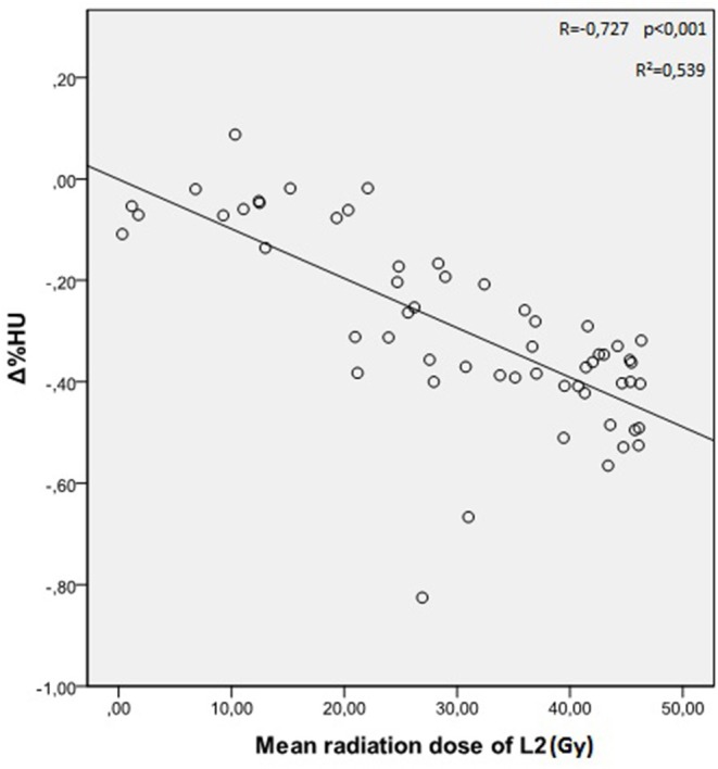
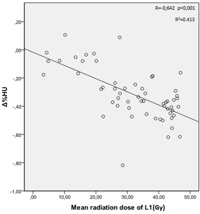
We aimed to evaluate osteoporosis, bone mineral density, and fracture risk in irradiated patients by computerized tomography derived Hounsfield Units (HUs) calculated from radiation treatment planning system. Fifty-seven patients operated for gastric adenocarcinoma who received adjuvant abdominal radiotherapy were included in the study group. Thirty-four patients who were not irradiated after surgery comprised the control group. HUs of T12, L1, L2 vertebral bodies were measured from the computerized tomographies imported to the treatment planning system for all the patients. While the measurements were obtained just after surgery and 1 year later after surgery in the control group, the same measurements were obtained just before irradiation and 1 year after radiotherapy in the study group. Percent change in HU values (Δ%HU) was determined for each group. Vertebral compression fractures, which are the consequence of radiation induced osteoporosis and bone toxicity were assessed during follow-up. There was no statistical significant difference in HU values measured for all the vertebrae between the study and the control group at the onset of the study. While HU values decreased significantly in the study group, there was no significant reduction in HU values in the control group after 1 year. significant correlation was found between Δ%HU and the radiation dose received by each vertebra. Insufficiency fractures (IFs) were observed only in the irradiated patients (4 out of 57 patients) with the cumulative incidence of 7%. HU values are very valuable in determining bone mineral density and fracture risk. Radiation treatment planning system can be utilized to determine HU values. IFs are common after abdominal radiotherapy in patients with low vertebral HU values detected during radiation treatment planning. Radiation dose to the vertebral bones with low HU values should be limited below 20 Gy to prevent late radiation related bone toxicity.

摘要

我们旨在通过从放射治疗计划系统计算得出的计算机断层扫描亨氏单位(HU)来评估接受放疗患者的骨质疏松症、骨密度和骨折风险。研究组纳入了57例接受辅助腹部放疗的胃腺癌手术患者。34例术后未接受放疗的患者组成对照组。对所有患者,从导入治疗计划系统的计算机断层扫描中测量T12、L1、L2椎体的HU值。对照组在术后即刻和术后1年进行测量,而研究组在放疗前即刻和放疗后1年进行相同测量。确定每组HU值的百分比变化(Δ%HU)。在随访期间评估作为放射诱导骨质疏松症和骨毒性后果的椎体压缩骨折。研究开始时,研究组和对照组所有椎体测量的HU值无统计学显著差异。研究组HU值显著下降,而对照组1年后HU值无显著降低。发现Δ%HU与每个椎体接受的放射剂量之间存在显著相关性。仅在接受放疗的患者(57例患者中有4例)中观察到骨质疏松性骨折(IFs),累积发生率为7%。HU值在确定骨密度和骨折风险方面非常有价值。放射治疗计划系统可用于确定HU值。在放射治疗计划期间检测到椎体HU值低的患者中,腹部放疗后IFs很常见。应将低HU值椎体的放射剂量限制在20 Gy以下,以预防晚期放射相关骨毒性。

https://cdn.ncbi.nlm.nih.gov/pmc/blobs/f6d6/7237579/ab0c27416c79/fonc-10-00742-g0003.jpg
https://cdn.ncbi.nlm.nih.gov/pmc/blobs/f6d6/7237579/ada7dbe5e66e/fonc-10-00742-g0001.jpg
https://cdn.ncbi.nlm.nih.gov/pmc/blobs/f6d6/7237579/044cc1ed33b6/fonc-10-00742-g0002.jpg
https://cdn.ncbi.nlm.nih.gov/pmc/blobs/f6d6/7237579/ab0c27416c79/fonc-10-00742-g0003.jpg
https://cdn.ncbi.nlm.nih.gov/pmc/blobs/f6d6/7237579/ada7dbe5e66e/fonc-10-00742-g0001.jpg
https://cdn.ncbi.nlm.nih.gov/pmc/blobs/f6d6/7237579/044cc1ed33b6/fonc-10-00742-g0002.jpg
https://cdn.ncbi.nlm.nih.gov/pmc/blobs/f6d6/7237579/ab0c27416c79/fonc-10-00742-g0003.jpg

相似文献

1
CT Derived Hounsfield Unit: An Easy Way to Determine Osteoporosis and Radiation Related Fracture Risk in Irradiated Patients.CT衍生的亨氏单位:一种确定受辐照患者骨质疏松症和辐射相关骨折风险的简便方法。
Front Oncol. 2020 May 13;10:742. doi: 10.3389/fonc.2020.00742. eCollection 2020.
2
Bone mineral density loss in thoracic and lumbar vertebrae following radiation for abdominal cancers.腹部癌症放疗后胸腰椎骨密度丢失。
Radiother Oncol. 2016 Mar;118(3):430-6. doi: 10.1016/j.radonc.2016.03.002. Epub 2016 Mar 15.
3
Opportunistic use of chest low-dose computed tomography (LDCT) imaging for low bone mineral density and osteoporosis screening: cutoff thresholds for the attenuation values of the lower thoracic and upper lumbar vertebrae.胸部低剂量计算机断层扫描(LDCT)成像在低骨密度和骨质疏松症筛查中的机会性应用:下胸椎和上腰椎衰减值的截断阈值
Quant Imaging Med Surg. 2024 Jul 1;14(7):4792-4803. doi: 10.21037/qims-24-59. Epub 2024 Jun 24.
4
What is the role of CT-based Hounsfield unit assessment in the evaluation of bone mineral density in patients undergoing 1- or 2-level lumbar spinal fusion for degenerative spinal pathologies? A prospective study.CT 基于亨氏单位评估在评估因退行性脊柱病变行 1 或 2 节段腰椎融合术患者的骨密度中的作用?一项前瞻性研究。
Spine J. 2023 Oct;23(10):1427-1434. doi: 10.1016/j.spinee.2023.05.015. Epub 2023 Jun 2.
5
Osteoporosis development and vertebral fractures after abdominal irradiation in patients with gastric cancer.胃癌患者腹部放疗后骨质疏松症的发展和椎体骨折。
BMC Cancer. 2018 Oct 11;18(1):972. doi: 10.1186/s12885-018-4899-z.
6
Dual-Energy X-Ray Absorptiometry (DEXA) Scan Versus Computed Tomography for Bone Density Assessment.双能X线吸收法(DEXA)扫描与计算机断层扫描用于骨密度评估的比较
Cureus. 2021 Feb 10;13(2):e13261. doi: 10.7759/cureus.13261.
7
Assessing the effects of lumbar posterior stabilization and fusion to vertebral bone density in stabilized and adjacent segments by using Hounsfield unit.利用亨氏单位评估腰椎后路稳定及融合对稳定节段和相邻节段椎体骨密度的影响。
J Spine Surg. 2017 Dec;3(4):548-553. doi: 10.21037/jss.2017.09.05.
8
Diagnostic efficacy of routine contrast-enhanced abdominal CT for the assessment of osteoporosis in the Turkish population.常规增强腹部 CT 诊断效能评估土耳其人群骨质疏松症。
Turk J Med Sci. 2020 Feb 13;50(1):110-116. doi: 10.3906/sag-1906-34.
9
Hounsfield unit for assessing bone mineral density distribution within lumbar vertebrae and its clinical values.用于评估腰椎骨矿物质密度分布的亨氏单位及其临床价值。
Front Endocrinol (Lausanne). 2024 Jun 13;15:1398367. doi: 10.3389/fendo.2024.1398367. eCollection 2024.
10
Value of Hounsfield units measured by chest computed tomography for assessing bone density in the thoracolumbar segment of the thoracic spine.胸部计算机断层扫描测量的亨氏单位在评估胸椎胸腰段骨密度中的价值。
Asian Spine J. 2024 Jun;18(3):336-345. doi: 10.31616/asj.2023.0438. Epub 2024 Jun 25.

引用本文的文献

1
Risk factors for radiation-induced rib fractures following proton beam therapy for stage I non-small cell lung cancer: a retrospective study.I期非小细胞肺癌质子束治疗后放射性肋骨骨折的危险因素:一项回顾性研究。
BMC Cancer. 2025 Apr 14;25(1):682. doi: 10.1186/s12885-025-14047-6.
2
The Incidence of Osteoporosis in Hepatocellular Carcinoma Patients Under 65: A Retrospective Cohort Study.65岁以下肝细胞癌患者骨质疏松症的发病率:一项回顾性队列研究
Cureus. 2025 Mar 12;17(3):e80490. doi: 10.7759/cureus.80490. eCollection 2025 Mar.
3
Hounsfield units predict vertebral compression fractures in gastric cancer survivors after adjuvant irradiation.

本文引用的文献

1
Osteoporosis development and vertebral fractures after abdominal irradiation in patients with gastric cancer.胃癌患者腹部放疗后骨质疏松症的发展和椎体骨折。
BMC Cancer. 2018 Oct 11;18(1):972. doi: 10.1186/s12885-018-4899-z.
2
Opportunistic screening for osteoporosis by routine CT in Southern Europe.在南欧,通过常规 CT 进行骨质疏松症的机会性筛查。
Osteoporos Int. 2017 Mar;28(3):983-990. doi: 10.1007/s00198-016-3804-3. Epub 2017 Jan 20.
3
Bone mineral density loss in thoracic and lumbar vertebrae following radiation for abdominal cancers.
亨斯菲尔德单位可预测胃癌幸存者辅助放疗后椎体压缩骨折。
Radiat Oncol J. 2025 Mar;43(1):30-39. doi: 10.3857/roj.2024.00409. Epub 2025 Feb 26.
4
Osteoporosis is not associated with increased reoperation or fracture risk three years after total ankle arthroplasty: a retrospective cohort study.全踝关节置换术后三年,骨质疏松与再次手术或骨折风险增加无关:一项回顾性队列研究。
Osteoporos Int. 2025 May;36(5):907-916. doi: 10.1007/s00198-025-07473-6. Epub 2025 Apr 4.
5
Monte Carlo modeling of radiation dose from radiation therapy with superficial x-rays.浅表X射线放射治疗辐射剂量的蒙特卡罗模拟
J Appl Clin Med Phys. 2025 Jun;26(6):e70062. doi: 10.1002/acm2.70062. Epub 2025 Mar 4.
6
Radiomic-based biomarkers: Transforming age and body composition metrics into personalized age-informed indices.基于影像组学的生物标志物:将年龄和身体成分指标转化为个性化的年龄相关指数。
Clin Transl Sci. 2024 Dec;17(12):e70062. doi: 10.1111/cts.70062.
7
The variability of CT scan protocols for total hip arthroplasty: a call for harmonisation.全髋关节置换术CT扫描协议的变异性:呼吁统一
EFORT Open Rev. 2023 Nov 1;8(11):809-817. doi: 10.1530/EOR-22-0141.
8
Computed Tomography Attenuation of Three-Dimensional (3D) Printing Materials-Depository to Aid in Constructing 3D-Printed Phantoms.用于辅助构建3D打印体模的三维(3D)打印材料储存库的计算机断层扫描衰减
Micromachines (Basel). 2023 Oct 14;14(10):1928. doi: 10.3390/mi14101928.
9
Smoking index and COPD duration as potential risk factors for development of osteoporosis in patients with non-small cell lung cancer - A retrospective case control study evaluated by CT Hounsfield unit.吸烟指数和慢性阻塞性肺疾病(COPD)病程作为非小细胞肺癌患者骨质疏松症发生的潜在危险因素——一项通过CT亨氏单位评估的回顾性病例对照研究
Heliyon. 2023 Oct 10;9(10):e20885. doi: 10.1016/j.heliyon.2023.e20885. eCollection 2023 Oct.
10
Association of radiotherapy with thoracic vertebral fractures in esophageal squamous cell carcinoma: A retrospective cohort study.放疗与食管鳞癌胸腰椎骨折的相关性:一项回顾性队列研究。
Medicine (Baltimore). 2023 Sep 29;102(39):e35304. doi: 10.1097/MD.0000000000035304.
腹部癌症放疗后胸腰椎骨密度丢失。
Radiother Oncol. 2016 Mar;118(3):430-6. doi: 10.1016/j.radonc.2016.03.002. Epub 2016 Mar 15.
4
Vertebral compression fractures after stereotactic body radiation therapy: a large, multi-institutional, multinational evaluation.立体定向体部放射治疗后椎体压缩骨折:一项大型、多机构、跨国评估。
J Neurosurg Spine. 2016 Jun;24(6):928-36. doi: 10.3171/2015.10.SPINE141261. Epub 2016 Feb 19.
5
Bone Health and Pelvic Radiotherapy.骨骼健康与盆腔放疗
Clin Oncol (R Coll Radiol). 2015 Nov;27(11):668-78. doi: 10.1016/j.clon.2015.07.006. Epub 2015 Aug 11.
6
Opportunistic screening for osteoporosis on routine computed tomography? An external validation study.在常规计算机断层扫描中对骨质疏松症进行机会性筛查?一项外部验证研究。
Eur Radiol. 2015 Jul;25(7):2074-9. doi: 10.1007/s00330-014-3584-0. Epub 2015 Jan 17.
7
Insufficiency fracture after radiation therapy.放射治疗后发生的不全骨折。
Radiat Oncol J. 2014 Dec;32(4):213-20. doi: 10.3857/roj.2014.32.4.213. Epub 2014 Dec 30.
8
The use of computed tomography attenuation to evaluate osteoporosis following acute fractures of the thoracic and lumbar vertebra.利用计算机断层扫描衰减评估胸腰椎急性骨折后的骨质疏松情况。
Geriatr Orthop Surg Rehabil. 2014 Jun;5(2):50-5. doi: 10.1177/2151458514525042.
9
Androgen-deprivation-associated bone disease.雄激素剥夺相关骨病
Curr Opin Urol. 2014 Nov;24(6):601-7. doi: 10.1097/MOU.0000000000000101.
10
Use of computed tomography for assessing bone mineral density.使用计算机断层扫描评估骨密度。
Neurosurg Focus. 2014;37(1):E4. doi: 10.3171/2014.5.FOCUS1483.