• 文献检索
  • 文档翻译
  • 深度研究
  • 学术资讯
  • Suppr Zotero 插件Zotero 插件
  • 邀请有礼
  • 套餐&价格
  • 历史记录
应用&插件
Suppr Zotero 插件Zotero 插件浏览器插件Mac 客户端Windows 客户端微信小程序
定价
高级版会员购买积分包购买API积分包
服务
文献检索文档翻译深度研究API 文档MCP 服务
关于我们
关于 Suppr公司介绍联系我们用户协议隐私条款
关注我们

Suppr 超能文献

核心技术专利:CN118964589B侵权必究
粤ICP备2023148730 号-1Suppr @ 2026

文献检索

告别复杂PubMed语法,用中文像聊天一样搜索,搜遍4000万医学文献。AI智能推荐,让科研检索更轻松。

立即免费搜索

文件翻译

保留排版,准确专业,支持PDF/Word/PPT等文件格式,支持 12+语言互译。

免费翻译文档

深度研究

AI帮你快速写综述,25分钟生成高质量综述,智能提取关键信息,辅助科研写作。

立即免费体验

更快、更锐利、更精确:自动化簇-FLIM 在临床前测试中直接识别治疗诊断在活细胞和组织中的细胞内命运。

Faster, sharper, more precise: Automated Cluster-FLIM in preclinical testing directly identifies the intracellular fate of theranostics in live cells and tissue.

机构信息

Institute of Experimental Physics, Freie Universität Berlin, Arnimallee 14, 14195 Berlin, Germany.

Institute of Pharmacy (Pharmacology and Toxicology), Freie Universität Berlin, Königin-Luise-Str. 2+4, 14195 Berlin, Germany.

出版信息

Theranostics. 2020 May 15;10(14):6322-6336. doi: 10.7150/thno.42581. eCollection 2020.

DOI:10.7150/thno.42581
PMID:32483455
原文链接:https://pmc.ncbi.nlm.nih.gov/articles/PMC7255044/
Abstract

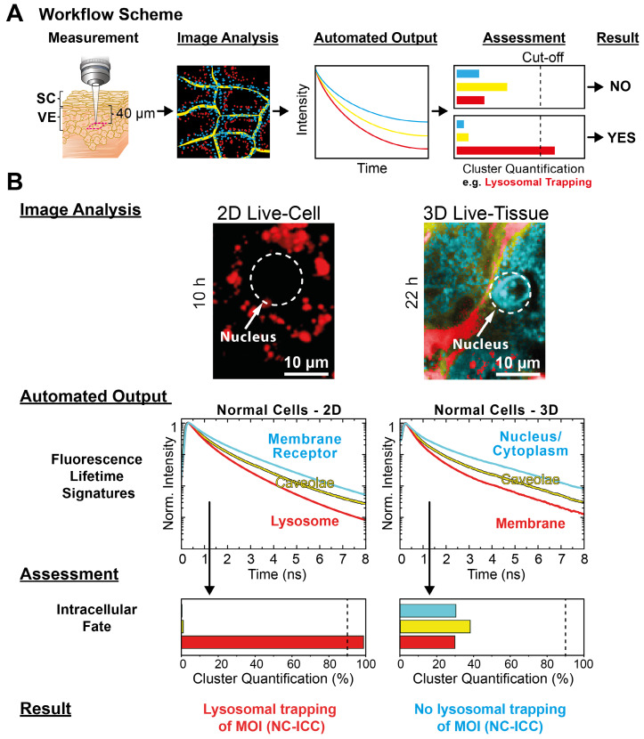
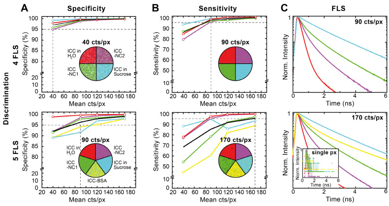
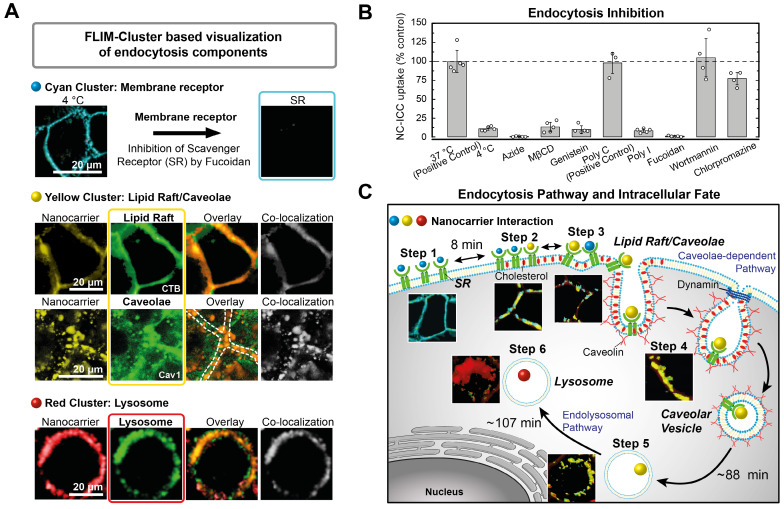
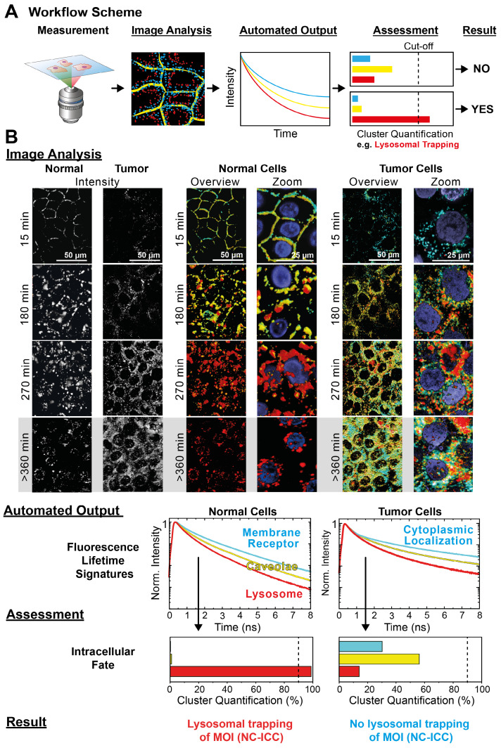
Fluorescence microscopy is widely used for high content screening in 2D cell cultures and 3D models. In particular, 3D tissue models are gaining major relevance in modern drug development. Enabling direct multiparametric evaluation of complex samples, fluorescence lifetime imaging (FLIM) adds a further level to intensity imaging by the sensitivity of the fluorescence lifetime to the microenvironment. However, the use of FLIM is limited amongst others by the acquisition of sufficient photon numbers without phototoxic effects in live cells. Herein, we developed a new cluster-based analysis method to enhance insight, and significantly speed up analysis and measurement time for the accurate translation of fluorescence lifetime information into pharmacological pathways. : We applied a fluorescently-labeled dendritic core-multishell nanocarrier and its cargo Bodipy as molecules of interest (MOI) to human cells and reconstructed human tissue. Following the sensitivity and specificity assessment of the fitting-free Cluster-FLIM analysis of data and , we evaluated the dynamics of cellular molecule uptake and intracellular interactions. For 3D live tissue investigations, we applied multiphoton (mp) FLIM. Owing to Cluster-FLIM's statistics-based fitting-free analysis, we utilized this approach for automatization. : To discriminate the fluorescence lifetime signatures of 5 different fluorescence species in a single color channel, the Cluster-FLIM method requires only 170, respectively, 90 counts per pixel to obtain 95% sensitivity (hit rate) and 95% specificity (correct rejection rate). Cluster-FLIM revealed cellular interactions of MOIs, representing their spatiotemporal intracellular fate. In a setting of an automated workflow, the assessment of lysosomal trapping of the MOI revealed relevant differences between normal and tumor cells, as well as between 2D and 3D models. : The automated Cluster-FLIM tool is fitting-free, providing images with enhanced information, contrast, and spatial resolution at short exposure times and low fluorophore concentrations. Thereby, Cluster-FLIM increases the applicability of FLIM in high content analysis of target molecules in drug development and beyond.

摘要

荧光显微镜广泛应用于 2D 细胞培养物和 3D 模型的高通量筛选。特别是,3D 组织模型在现代药物开发中具有重要意义。荧光寿命成像(FLIM)通过荧光寿命对微环境的敏感性,使复杂样品的直接多参数评估成为可能,为强度成像增加了一个新的层次。然而,FLIM 的应用受到限制,例如在活细胞中没有光毒性作用的情况下获取足够的光子数。在此,我们开发了一种新的基于聚类的分析方法,以提高对荧光寿命信息转化为药理途径的深入了解,并显著加快分析和测量时间。

我们应用了一种荧光标记的树突状核壳纳米载体及其作为感兴趣分子(MOI)的货物 Bodipy 到人类细胞和重建的人类组织中。在对无拟合聚类-FLIM 数据分析的灵敏度和特异性进行评估后,我们评估了细胞内分子摄取和细胞内相互作用的动力学。对于 3D 活组织研究,我们应用了多光子(mp)FLIM。由于 Cluster-FLIM 基于统计学的无拟合分析,我们利用这种方法实现了自动化。

为了在单个颜色通道中区分 5 种不同荧光物质的荧光寿命特征,Cluster-FLIM 方法仅需要 170 个,分别为 90 个像素/计数,以获得 95%的灵敏度(命中率)和 95%的特异性(正确拒绝率)。Cluster-FLIM 揭示了 MOI 的细胞相互作用,代表了它们的时空细胞内命运。在自动化工作流程的设置中,对 MOI 溶酶体捕获的评估揭示了正常细胞和肿瘤细胞之间以及 2D 和 3D 模型之间的相关差异。

自动聚类-FLIM 工具是无拟合的,在短曝光时间和低荧光浓度下提供具有增强信息、对比度和空间分辨率的图像。因此,Cluster-FLIM 提高了 FLIM 在药物开发和其他领域中对靶分子高通量分析的适用性。

https://cdn.ncbi.nlm.nih.gov/pmc/blobs/91df/7255044/6663f67f5879/thnov10p6322g006.jpg
https://cdn.ncbi.nlm.nih.gov/pmc/blobs/91df/7255044/000ceb02a6ba/thnov10p6322g001.jpg
https://cdn.ncbi.nlm.nih.gov/pmc/blobs/91df/7255044/367f07686ad7/thnov10p6322g002.jpg
https://cdn.ncbi.nlm.nih.gov/pmc/blobs/91df/7255044/7ba40be68790/thnov10p6322g003.jpg
https://cdn.ncbi.nlm.nih.gov/pmc/blobs/91df/7255044/d5f0d400c64a/thnov10p6322g004.jpg
https://cdn.ncbi.nlm.nih.gov/pmc/blobs/91df/7255044/3f43d0069d54/thnov10p6322g005.jpg
https://cdn.ncbi.nlm.nih.gov/pmc/blobs/91df/7255044/6663f67f5879/thnov10p6322g006.jpg

相似文献

1
Faster, sharper, more precise: Automated Cluster-FLIM in preclinical testing directly identifies the intracellular fate of theranostics in live cells and tissue.更快、更锐利、更精确:自动化簇-FLIM 在临床前测试中直接识别治疗诊断在活细胞和组织中的细胞内命运。
Theranostics. 2020 May 15;10(14):6322-6336. doi: 10.7150/thno.42581. eCollection 2020.
2
Image restoration for fluorescence lifetime imaging microscopy (FLIM).用于荧光寿命成像显微镜(FLIM)的图像恢复
Opt Express. 2008 Nov 10;16(23):19192-200. doi: 10.1364/oe.16.019192.
3
Multiphoton FLIM imaging of NAD(P)H and FAD with one excitation wavelength.利用单一激发波长的多光子 FLIM 成像技术对 NAD(P)H 和 FAD 进行成像。
J Biomed Opt. 2020 Jan;25(1):1-16. doi: 10.1117/1.JBO.25.1.014510.
4
Time-resolved fluorescence microscopy (FLIM) as an analytical tool in skin nanomedicine.时间分辨荧光显微镜(FLIM)作为皮肤纳米医学中的一种分析工具。
Eur J Pharm Biopharm. 2017 Jul;116:111-124. doi: 10.1016/j.ejpb.2017.01.005. Epub 2017 Jan 21.
5
Fluorescence lifetime microscopy of tumor cell invasion, drug delivery, and cytotoxicity.肿瘤细胞侵袭、药物递送及细胞毒性的荧光寿命显微镜检查
Methods Enzymol. 2012;504:109-25. doi: 10.1016/B978-0-12-391857-4.00005-7.
6
Fluorescence lifetime imaging microscopy for quantitative biological imaging.用于定量生物成像的荧光寿命成像显微镜。
Methods Cell Biol. 2013;114:457-88. doi: 10.1016/B978-0-12-407761-4.00020-8.
7
Two-photon fluorescence lifetime imaging of intrinsic NADH in three-dimensional tumor models.三维肿瘤模型中内源性 NADH 的双光子荧光寿命成像。
Cytometry A. 2019 Jan;95(1):80-92. doi: 10.1002/cyto.a.23632. Epub 2018 Oct 21.
8
Light-field tomographic fluorescence lifetime imaging microscopy.光场层析荧光寿命成像显微镜。
Proc Natl Acad Sci U S A. 2024 Oct;121(40):e2402556121. doi: 10.1073/pnas.2402556121. Epub 2024 Sep 25.
9
Autofluorescence lifetime imaging of cellular metabolism: Sensitivity toward cell density, pH, intracellular, and intercellular heterogeneity.细胞代谢的自体荧光寿命成像:对细胞密度、pH 值、细胞内和细胞间异质性的敏感性。
Cytometry A. 2019 Jan;95(1):56-69. doi: 10.1002/cyto.a.23603. Epub 2018 Oct 8.
10
Single-photon peak event detection (SPEED): a computational method for fast photon counting in fluorescence lifetime imaging microscopy.单光子峰事件检测 (SPEED):一种用于荧光寿命成像显微镜中快速光子计数的计算方法。
Opt Express. 2021 Nov 8;29(23):37759-37775. doi: 10.1364/OE.439675.

引用本文的文献

1
Fluorescence Lifetime Measurements and Analyses: Protocols Using Flow Cytometry and High-Throughput Microscopy.荧光寿命测量和分析:流式细胞术和高通量显微镜的使用方案。
Methods Mol Biol. 2024;2779:323-351. doi: 10.1007/978-1-0716-3738-8_15.
2
Visualization of Nanocarriers and Drugs in Cells and Tissue.纳米载体和药物在细胞和组织中的可视化。
Handb Exp Pharmacol. 2024;284:153-189. doi: 10.1007/164_2023_684.
3
QuasAr Odyssey: the origin of fluorescence and its voltage sensitivity in microbial rhodopsins.QuasAr Odyssey:微生物视紫红质中荧光的起源及其对电压的敏感性。

本文引用的文献

1
Label-free whole-colony imaging and metabolic analysis of metastatic pancreatic cancer by an autoregulating flexible optical system.无标记全菌落成像和代谢分析转移性胰腺癌的自调节柔性光学系统。
Theranostics. 2020 Jan 1;10(4):1849-1860. doi: 10.7150/thno.40869. eCollection 2020.
2
Imaging-assisted anticancer nanotherapy.影像辅助抗癌纳米治疗。
Theranostics. 2020 Jan 1;10(3):956-967. doi: 10.7150/thno.38288. eCollection 2020.
3
Lightsheet fluorescence lifetime imaging microscopy with wide-field time-correlated single photon counting.
Nat Commun. 2022 Sep 20;13(1):5501. doi: 10.1038/s41467-022-33084-4.
4
In Vitro Models of Biological Barriers for Nanomedical Research.用于纳米医学研究的生物屏障的体外模型。
Int J Mol Sci. 2022 Aug 10;23(16):8910. doi: 10.3390/ijms23168910.
5
Comparison of fluorescence lifetime and multispectral imaging for quantitative multiplexing in biological tissue.用于生物组织定量多重分析的荧光寿命与多光谱成像比较
Biomed Opt Express. 2022 Jun 9;13(7):3854-3868. doi: 10.1364/BOE.459935. eCollection 2022 Jul 1.
6
Improved fluorescent phytochromes for in situ imaging.改良的荧光光光色素用于原位成像。
Sci Rep. 2022 Apr 4;12(1):5587. doi: 10.1038/s41598-022-09169-x.
7
Deep learning in macroscopic diffuse optical imaging.深度学习在宏观漫射光学成像中的应用。
J Biomed Opt. 2022 Feb;27(2). doi: 10.1117/1.JBO.27.2.020901.
8
Label-free sensing of cells with fluorescence lifetime imaging: The quest for metabolic heterogeneity.无标记细胞荧光寿命成像检测:探寻代谢异质性。
Proc Natl Acad Sci U S A. 2022 Mar 1;119(9). doi: 10.1073/pnas.2118241119.
9
Phasor-based image segmentation: machine learning clustering techniques.基于相量的图像分割:机器学习聚类技术。
Biomed Opt Express. 2021 May 17;12(6):3410-3422. doi: 10.1364/BOE.422766. eCollection 2021 Jun 1.
10
A Nomogram Based on a Collagen Feature Support Vector Machine for Predicting the Treatment Response to Neoadjuvant Chemoradiotherapy in Rectal Cancer Patients.基于胶原特征支持向量机的列线图预测直肠癌患者新辅助放化疗治疗反应
Ann Surg Oncol. 2021 Oct;28(11):6408-6421. doi: 10.1245/s10434-021-10218-4. Epub 2021 Jun 19.
基于宽场时间相关单光子计数的光片荧光寿命成像显微镜
J Biophotonics. 2020 Feb;13(2):e201960099. doi: 10.1002/jbio.201960099. Epub 2019 Nov 25.
4
Tumor microenvironment determines drug efficacy in vitro - apoptotic and anti-inflammatory effects of 15-lipoxygenase metabolite, 13-HpOTrE.肿瘤微环境决定体外药物疗效-15-脂氧合酶代谢物,13-HpODE 的促凋亡和抗炎作用。
Eur J Pharm Biopharm. 2019 Sep;142:1-7. doi: 10.1016/j.ejpb.2019.06.003. Epub 2019 Jun 6.
5
A deep learning-based algorithm for 2-D cell segmentation in microscopy images.基于深度学习的显微镜图像二维细胞分割算法。
BMC Bioinformatics. 2018 Oct 3;19(1):365. doi: 10.1186/s12859-018-2375-z.
6
FLIM reveals alternative EV-mediated cellular up-take pathways of paclitaxel.FLIM 揭示了紫杉醇的 EV 介导的细胞摄取的替代途径。
J Control Release. 2018 Aug 28;284:133-143. doi: 10.1016/j.jconrel.2018.06.015. Epub 2018 Jun 15.
7
Visualizing Oxidative Cellular Stress Induced by Nanoparticles in the Subcytotoxic Range Using Fluorescence Lifetime Imaging.使用荧光寿命成像技术可视化亚细胞毒性范围内纳米颗粒引起的氧化细胞应激。
Small. 2018 Jun;14(23):e1800310. doi: 10.1002/smll.201800310. Epub 2018 May 4.
8
Three-Dimensional Cell Culture Models in Drug Discovery and Drug Repositioning.药物发现与药物重新定位中的三维细胞培养模型
Front Pharmacol. 2018 Jan 23;9:6. doi: 10.3389/fphar.2018.00006. eCollection 2018.
9
Estimation of clinical trial success rates and related parameters.临床试验成功率及相关参数的估计。
Biostatistics. 2019 Apr 1;20(2):273-286. doi: 10.1093/biostatistics/kxx069.
10
Phasor-Fluorescence Lifetime Imaging Microscopy Analysis to Monitor Intercellular Drug Release from a pH-Sensitive Polymeric Nanocarrier.相衬荧光寿命成像显微镜分析监测 pH 敏感聚合物纳米载体的细胞间药物释放。
Anal Chem. 2018 Feb 6;90(3):2170-2177. doi: 10.1021/acs.analchem.7b04511. Epub 2018 Jan 25.