Discipline of Physiotherapy, Faculty of Medicine and Health, The University of Sydney, & The Northern Sydney Local Health District, The Kolling Research Institute, St. Leonards, New South Wales, Australia.
Physical Therapy and Human Movement Sciences, Feinberg School of Medicine, Northwestern University, Chicago, Illinois, United States of America.
PLoS One. 2020 Jun 2;15(6):e0234061. doi: 10.1371/journal.pone.0234061. eCollection 2020.
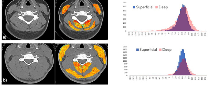
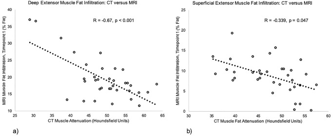
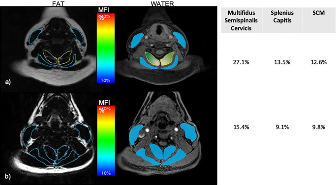
Here we present a secondary analysis from a parent database of 97 acutely injured participants enrolled in a prospective inception cohort study of whiplash recovery after motor vehicle collision (MVC). The purpose was to investigate the deep and superficial neck extensor muscles with peri-traumatic computed tomography (CT) and longitudinal measures of magnetic resonance imaging (MRI) in participants with varying levels of whiplash-related disability. Thirty-six underwent standard care imaging of the cervical spine with CT at a level-1 trauma designated emergency department. All 36 participants were assessed with MRI of the cervical spine at <1-week, 2-weeks, 3-, and 12-months post-injury and classified into three groups using initial pain severity and percentage scores on the Neck Disability Index (recovered (NDI of 0-8%), mild (NDI of 10-28%), or severe (NDI ≥ 30%)) at 3-months post MVC. CT muscle attenuation values were significantly correlated to muscle fat infiltration (MFI) on MRI at one-week post MVC. There was no significant difference in muscle attenuation across groups at the time of enrollment. A trend of lower muscle attenuation in the deep compared to the superficial extensors was observed in the severe group. MFI values in the deep muscles on MRI were significantly higher in the severe group when compared to the mild group at 1-year post MVC. This study provides further evidence that the magnitude of 1) deep MFI appears unique to those at risk of and eventually transitioning to chronic WAD and that 2) pre- or peri-traumatic muscular health, determined by CT muscle attenuation, may be contribute to our understanding of long-term recovery.
在这里,我们呈现了一项来自母体数据库的二次分析,该数据库纳入了 97 名急性创伤参与者,这些参与者参与了机动车碰撞后颈痛恢复的前瞻性队列研究。目的是研究深层和浅层颈伸肌在创伤后计算机断层扫描(CT)和磁共振成像(MRI)的纵向测量中,在颈痛相关残疾程度不同的参与者中的情况。36 名参与者在 1 级创伤指定急诊室接受了颈椎 CT 标准护理成像。所有 36 名参与者均在损伤后 <1 周、2 周、3 个月和 12 个月接受了颈椎 MRI 评估,并根据初始疼痛严重程度和颈痛障碍指数(NDI)的百分比评分(恢复组(NDI 为 0-8%)、轻度组(NDI 为 10-28%)或重度组(NDI≥30%))在 MVC 后 3 个月进行分类。CT 肌肉衰减值与损伤后 1 周的 MRI 肌肉脂肪浸润(MFI)显著相关。在入组时,各组之间的肌肉衰减值没有显著差异。在重度组中,与浅层伸肌相比,深层肌肉的衰减值较低。与轻度组相比,重度组在 MVC 后 1 年的 MRI 上深层肌肉的 MFI 值显著更高。这项研究进一步证明了 1)深层 MFI 的严重程度似乎是那些有发展为慢性 WAD 风险并最终过渡到慢性 WAD 的人的独特特征,2)CT 肌肉衰减值确定的受伤前或受伤时的肌肉健康状况可能有助于我们理解长期恢复。