Fundación Oftalmológica Los Andes, Vitacura, Santiago de Chile, Chile.
Department of Ophthalmology, Japan Community Healthcare Organization, Chukyo Hospital, Nagoya, Aichi, Japan.
Asia Pac J Ophthalmol (Phila). 2020 May-Jun;9(3):269-277. doi: 10.1097/APO.0000000000000292.
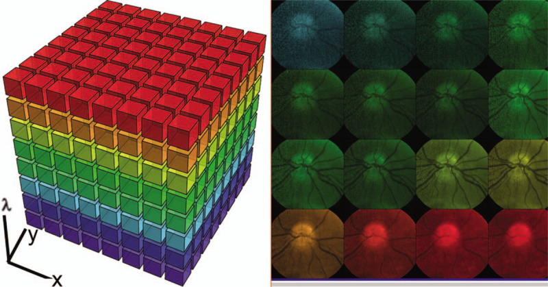
The diagnosis and treatment of medical retinal disease is now inseparable from retinal imaging in all its multimodal incarnations. The purpose of this article is to present a selection of very different retinal imaging techniques that are truly translational, in the sense that they are not only new, but can guide us to new understandings of disease processes or interventions that are not accessible by present methods. Quantitative autofluorescence imaging, now available for clinical investigation, has already fundamentally changed our understanding of the role of lipofuscin in age-related macular degeneration. Hyperspectral autofluorescence imaging is bench science poised not only to unravel the molecular basis of retinal pigment epithelium fluorescence, but also to be translated into a clinical camera for earliest detection of age-related macular degeneration. The ophthalmic endoscope for vitreous surgery is a radically new retinal imaging system that enables surgical approaches heretofore impossible while it captures subretinal images of living tissue. Remote retinal imaging coupled with deep learning artificial intelligence will transform the very fabric of future medical care.
医学视网膜疾病的诊断和治疗现在离不开各种模态的视网膜成像。本文的目的是介绍一系列非常不同的真正具有转化意义的视网膜成像技术,这些技术不仅是新的,而且可以引导我们对疾病过程或现有方法无法获得的干预措施有新的认识。定量自发荧光成像现在可用于临床研究,它已经从根本上改变了我们对脂褐素在年龄相关性黄斑变性中的作用的理解。高光谱自发荧光成像处于基础科学阶段,不仅可以揭示视网膜色素上皮荧光的分子基础,而且可以转化为临床相机,用于最早发现年龄相关性黄斑变性。玻璃体手术用眼科内窥镜是一种全新的视网膜成像系统,它不仅可以揭示视网膜色素上皮荧光的分子基础,而且可以转化为临床相机,用于最早发现年龄相关性黄斑变性。用于玻璃体手术的眼科内窥镜是一种全新的视网膜成像系统,它不仅可以揭示视网膜色素上皮荧光的分子基础,而且可以转化为临床相机,用于最早发现年龄相关性黄斑变性。用于玻璃体手术的眼科内窥镜是一种全新的视网膜成像系统,它不仅可以揭示视网膜色素上皮荧光的分子基础,而且可以转化为临床相机,用于最早发现年龄相关性黄斑变性。它使以前不可能实现的手术方法成为可能,同时捕获活体组织的视网膜下图像。远程视网膜成像结合深度学习人工智能将彻底改变未来医疗保健的结构。