i3S - Instituto de Investigação e Inovação em Saúde, University of Porto, Rua Alfredo Allen 208, 4200-135, Porto, Portugal.
INEB - Instituto de Engenharia Biomédica, University of Porto, Rua Alfredo Allen 208, 4200-135, Porto, Portugal.
Cell Death Dis. 2020 Jun 2;11(6):415. doi: 10.1038/s41419-020-2626-6.
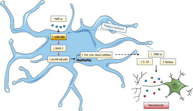
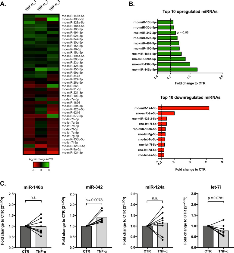
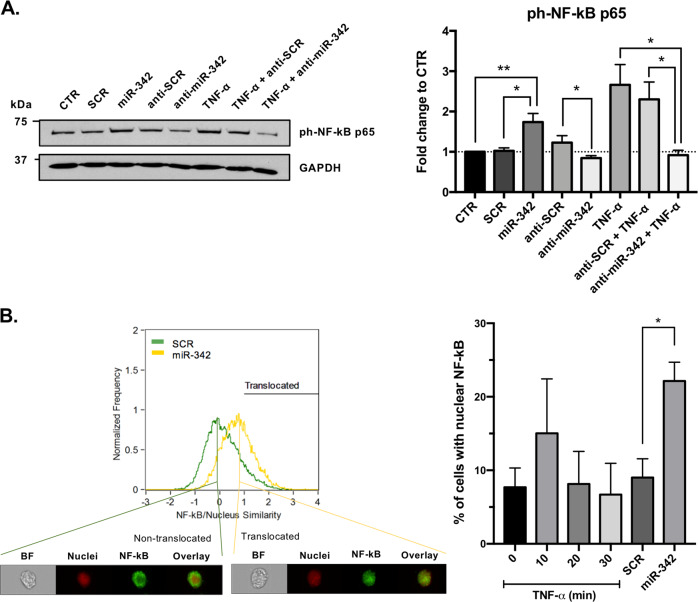
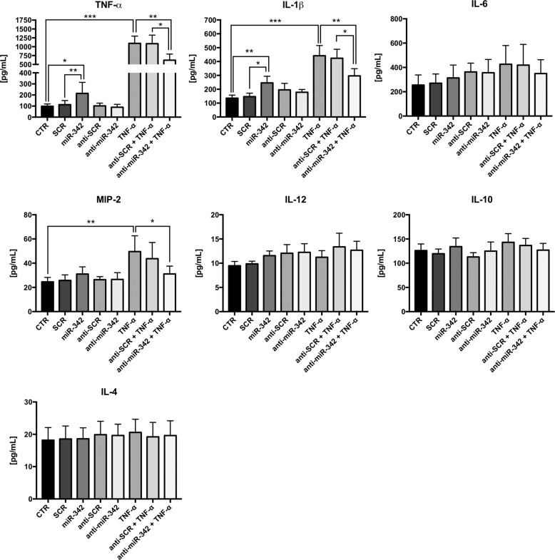
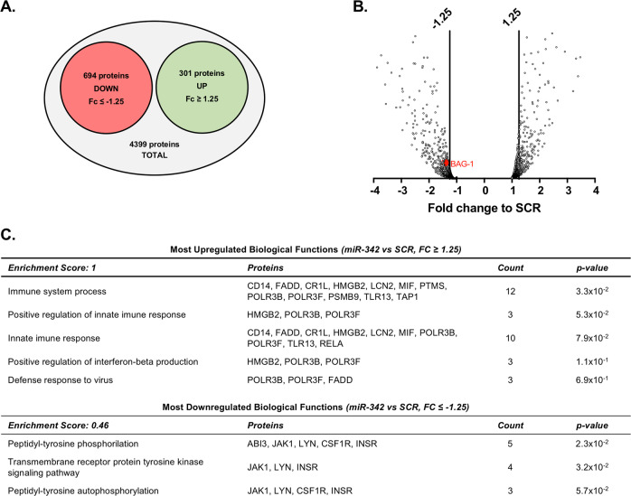
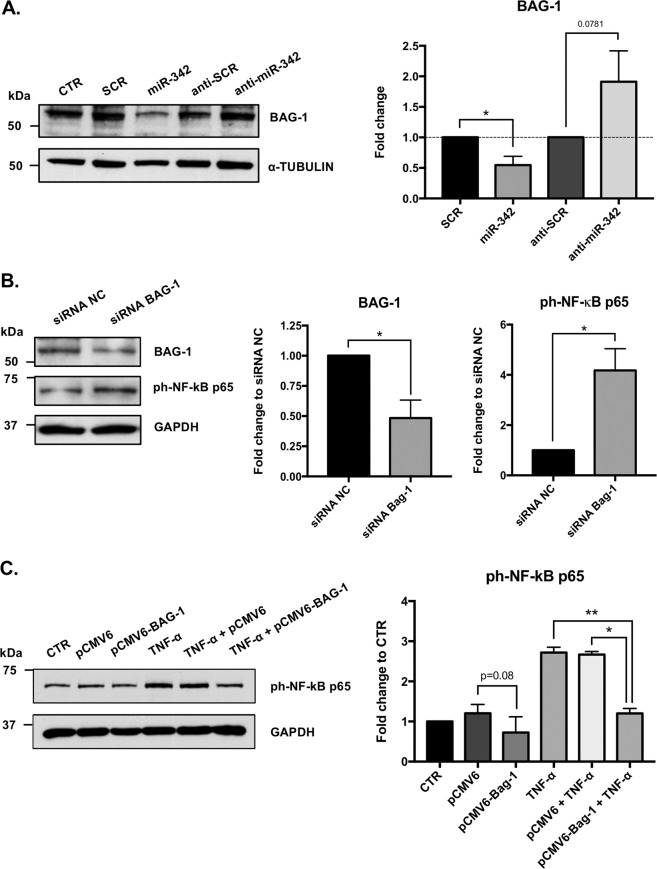
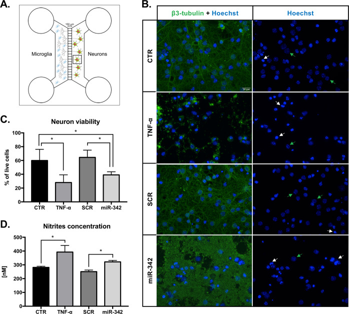
Growing evidences suggest that sustained neuroinflammation, caused by microglia overactivation, is implicated in the development and aggravation of several neurological and psychiatric disorders. In some pathological conditions, microglia produce increased levels of cytotoxic and inflammatory mediators, such as tumor necrosis factor alpha (TNF-α), which can reactivate microglia in a positive feedback mechanism. However, specific molecular mediators that can be effectively targeted to control TNF-α-mediated microglia overactivation, are yet to be uncovered. In this context, we aim to identify novel TNF-α-mediated micro(mi)RNAs and to dissect their roles in microglia activation, as well as to explore their impact on the cellular communication with neurons. A miRNA microarray, followed by RT-qPCR validation, was performed on TNF-α-stimulated primary rat microglia. Gain- and loss-of-function in vitro assays and proteomic analysis were used to dissect the role of miR-342 in microglia activation. Co-cultures of microglia with hippocampal neurons, using a microfluidic system, were performed to understand the impact on neurotoxicity. Stimulation of primary rat microglia with TNF-α led to an upregulation of Nos2, Tnf, and Il1b mRNAs. In addition, ph-NF-kB p65 levels were also increased. miRNA microarray analysis followed by RT-qPCR validation revealed that TNF-α stimulation induced the upregulation of miR-342. Interestingly, miR-342 overexpression in N9 microglia was sufficient to activate the NF-kB pathway by inhibiting BAG-1, leading to increased secretion of TNF-α and IL-1β. Conversely, miR-342 inhibition led to a strong decrease in the levels of these cytokines after TNF-α activation. In fact, both TNF-α-stimulated and miR-342-overexpressing microglia drastically affected neuron viability. Remarkably, increased levels of nitrites were detected in the supernatants of these co-cultures. Globally, our findings show that miR-342 is a crucial mediator of TNF-α-mediated microglia activation and a potential target to tackle microglia-driven neuroinflammation.
越来越多的证据表明,小胶质细胞过度激活引起的持续神经炎症与几种神经和精神疾病的发展和恶化有关。在某些病理条件下,小胶质细胞产生高水平的细胞毒性和炎症介质,如肿瘤坏死因子-α(TNF-α),这可以在正反馈机制中使小胶质细胞重新激活。然而,尚未发现可以有效靶向控制 TNF-α介导的小胶质细胞过度激活的特定分子介质。在这种情况下,我们旨在确定新的 TNF-α 介导的 micro(mi)RNAs,并剖析它们在小胶质细胞激活中的作用,以及探索它们对神经元与神经元之间细胞通讯的影响。对 TNF-α 刺激的原代大鼠小胶质细胞进行了 miRNA 微阵列分析,随后进行了 RT-qPCR 验证。体外获得和丧失功能试验以及蛋白质组学分析用于剖析 miR-342 在小胶质细胞激活中的作用。使用微流控系统对小胶质细胞与海马神经元进行共培养,以了解对神经毒性的影响。TNF-α刺激原代大鼠小胶质细胞导致 Nos2、Tnf 和 Il1b mRNA 的上调。此外,磷酸化 NF-kB p65 水平也增加。miRNA 微阵列分析随后进行 RT-qPCR 验证表明,TNF-α 刺激诱导了 miR-342 的上调。有趣的是,N9 小胶质细胞中 miR-342 的过表达足以通过抑制 BAG-1 激活 NF-kB 途径,导致 TNF-α 和 IL-1β 的分泌增加。相反,miR-342 的抑制导致 TNF-α 激活后这些细胞因子的水平大幅下降。事实上,TNF-α 刺激和 miR-342 过表达的小胶质细胞都严重影响了神经元的活力。值得注意的是,在这些共培养物的上清液中检测到亚硝酸盐水平升高。总体而言,我们的研究结果表明,miR-342 是 TNF-α 介导的小胶质细胞激活的关键介质,是治疗小胶质细胞驱动的神经炎症的潜在靶点。