Department of Exercise and Sport Science, University of North Carolina at Chapel Hill, Chapel Hill, NC, USA.
School of Sport and Exercise, University of Gloucestershire, Gloucester, UK.
Vasc Med. 2020 Oct;25(5):419-426. doi: 10.1177/1358863X20926588. Epub 2020 Jun 3.
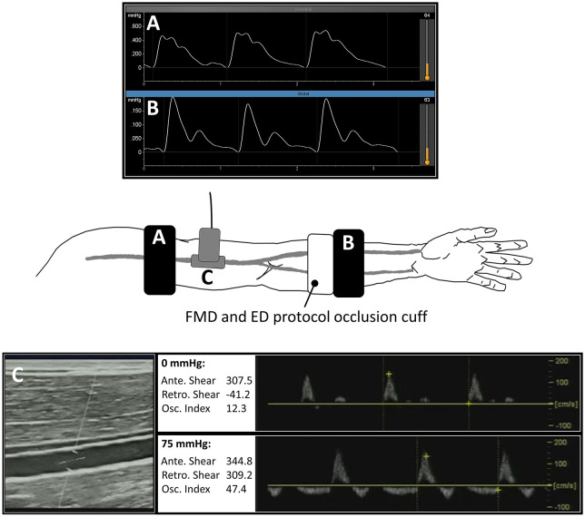
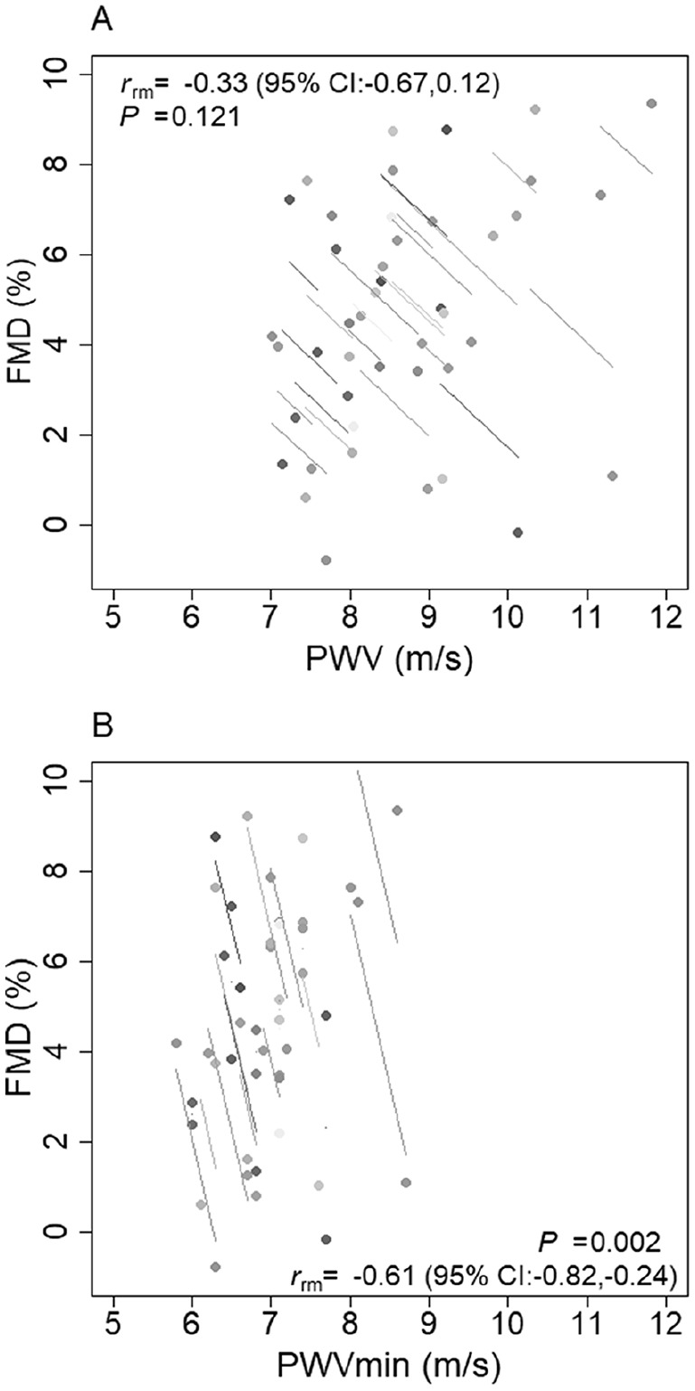
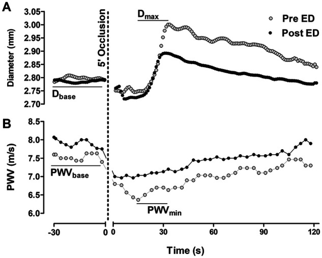
Flow-mediated slowing (FMS), defined as the minimum pulse wave velocity (PWV) during reactive hyperemia, is potentially a simple, user-objective test for examining endothelial function. The purpose of the current study was to determine the effects of a known endothelial dysfunction protocol on arm PWV and PWV. Complete data were successfully collected in 22 out of 23 healthy adults (23.8 years [SD 4.1], 16 F, 22.8 kg/m [SD 2.8]). Local endothelial dysfunction was induced by increasing retrograde shear stress in the upper arm, through inflation of a distal (forearm) tourniquet to 75 mmHg, for 30 min. Pre- and post-endothelial dysfunction, PWV was measured followed by simultaneous assessment of PWV and flow-mediated dilation (FMD). PWV was measured between the upper arm and wrist using an oscillometric device, and brachial FMD using ultrasound. FMD (%) and PWV (m/s) were calculated as the maximum increase in diameter and minimum PWV during reactive hyperemia, respectively. Endothelial dysfunction resulted in a large effect size (ES) decrease in FMD (∆ = -3.10%; 95% CI: -4.15, -2.05; ES = -1.3), and a moderate increase in PWV (∆ = 0.38 m/s; 95% CI: 0.07, 0.69; ES = 0.5) and PWV (∆ = 0.16 m/s; 95% CI: 0.05, 0.28; ES = 0.6). There was a large intra-individual (pre- vs post-endothelial dysfunction) association between FMD and PWV ( = -0.61; 95% CI: -0.82, -0.24). In conclusion, acute change in PWV and PWV are at least partially driven by changes in endothelial function.
血流介导的减速(FMS),定义为反应性充血期间的最小脉搏波速度(PWV),可能是一种简单的、用户客观的测试内皮功能的方法。本研究的目的是确定已知的内皮功能障碍方案对臂 PWV 和 PWV 的影响。在 23 名健康成年人中(23.8 岁[SD 4.1],16 名女性,22.8 kg/m[SD 2.8])成功收集了 22 名参与者的完整数据。通过将远侧(前臂)止血带充气至 75 mmHg,持续 30 分钟,在上臂中增加逆行剪切应力,从而诱导局部内皮功能障碍。在诱导内皮功能障碍前后,使用示波法测量 PWV,同时评估 PWV 和血流介导的扩张(FMD)。使用示波法设备在上臂和手腕之间测量 PWV,使用超声测量肱动脉 FMD。FMD(%)和 PWV(m/s)分别作为反应性充血期间直径的最大增加和最小 PWV 计算。内皮功能障碍导致 FMD 的大效应量(ES)降低(∆=-3.10%;95%CI:-4.15,-2.05;ES=-1.3),PWV 增加(∆=0.38 m/s;95%CI:0.07,0.69;ES=0.5)和 PWV(∆=0.16 m/s;95%CI:0.05,0.28;ES=0.6)适度增加。FMD 和 PWV 之间存在个体内(内皮功能障碍前后)的大关联(= -0.61;95%CI:-0.82,-0.24)。总之,PWV 和 PWV 的急性变化至少部分是由内皮功能的变化驱动的。