• 文献检索
  • 文档翻译
  • 深度研究
  • 学术资讯
  • Suppr Zotero 插件Zotero 插件
  • 邀请有礼
  • 套餐&价格
  • 历史记录
应用&插件
Suppr Zotero 插件Zotero 插件浏览器插件Mac 客户端Windows 客户端微信小程序
定价
高级版会员购买积分包购买API积分包
服务
文献检索文档翻译深度研究API 文档MCP 服务
关于我们
关于 Suppr公司介绍联系我们用户协议隐私条款
关注我们

Suppr 超能文献

核心技术专利:CN118964589B侵权必究
粤ICP备2023148730 号-1Suppr @ 2026

文献检索

告别复杂PubMed语法,用中文像聊天一样搜索,搜遍4000万医学文献。AI智能推荐,让科研检索更轻松。

立即免费搜索

文件翻译

保留排版,准确专业,支持PDF/Word/PPT等文件格式,支持 12+语言互译。

免费翻译文档

深度研究

AI帮你快速写综述,25分钟生成高质量综述,智能提取关键信息,辅助科研写作。

立即免费体验

马的关节沟模型中的 T2* 映射:观察胶原纤维方向的变化。

T2* mapping in an equine articular groove model: Visualizing changes in collagen orientation.

机构信息

Department of Radiology, University Medical Center Utrecht, Utrecht, The Netherlands.

Department of Clinical Sciences, Faculty of Veterinary Medicine, Utrecht University, Utrecht, The Netherlands.

出版信息

J Orthop Res. 2020 Nov;38(11):2383-2389. doi: 10.1002/jor.24764. Epub 2020 Jun 10.

DOI:10.1002/jor.24764
PMID:32492207
原文链接:https://pmc.ncbi.nlm.nih.gov/articles/PMC7687204/
Abstract

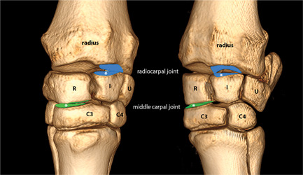
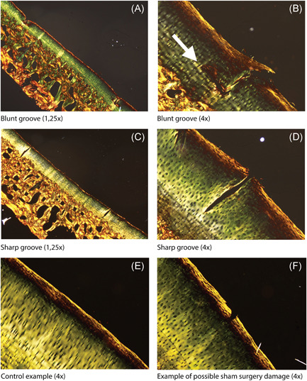
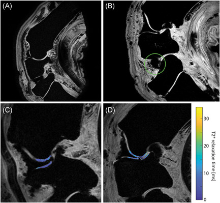
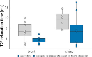
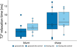
T2* mapping is promising for the evaluation of articular cartilage collagen. In this work, a groove model in a large animal is used as a model for posttraumatic arthritis. We hypothesized that T2* mapping could be employed to differentiate between healthy and (subtly) damaged cartilage. Eight carpal joints were obtained from four adult Shetland ponies that had been included in the groove study. In this model, grooves were surgically created on the proximal articular surface of the intermediate carpal bone (radiocarpal joint) and the radial facet of the third carpal bone (middle carpal joint) by either coarse disruption or sharp incision. After 9 months, T2* mapping of the entire carpal joint was carried out on a 7.0-T whole-body magnetic resonance imaging (MRI) scanner by means of a gradient echo multi-echo sequence. Afterwards, assessment of collagen orientation was carried out based on Picrosirius Red-stained histological sections, visualized by polarized light microscopy (PLM). The average T2* relaxation time in grooved samples was lower than in contralateral control sites. Opposite to the grooved areas, the "kissing sites" had a higher average T2* relaxation time than the grooved sites. PLM showed mild changes in orientation of the collagen fibers, particularly around blunt grooves. This work shows that T2* relaxation times are different in healthy cartilage vs (early) damaged cartilage, as induced by the equine groove model. Additionally, the average T2* relaxation times are different in kissing lesions vs the grooved sites.

摘要

T2* 映射有望用于评估关节软骨胶原。在这项工作中,使用大型动物的沟槽模型作为创伤后关节炎的模型。我们假设 T2* 映射可用于区分健康和(轻微)受损的软骨。从已纳入沟槽研究的四只成年设得兰矮种马中获得了八个腕关节。在该模型中,通过粗钝或锐切在中间腕骨(桡腕关节)的近侧关节面和第三腕骨(中间腕关节)的桡侧关节面上创建沟槽。9 个月后,在 7.0-T 全身磁共振成像(MRI)扫描仪上通过梯度回波多回波序列对整个腕关节进行 T2* 映射。之后,根据番红 O 染色的组织学切片,通过偏振光显微镜(PLM)进行胶原取向评估。有沟槽的样本的平均 T2* 弛豫时间低于对侧对照部位。与有沟槽的区域相反,“亲吻部位”的平均 T2* 弛豫时间高于有沟槽的部位。PLM 显示胶原纤维的取向有轻微变化,特别是在钝沟槽周围。这项工作表明,T2* 弛豫时间在健康软骨与(早期)受损软骨之间存在差异,这是由马的沟槽模型引起的。此外,亲吻病变与沟槽部位的平均 T2* 弛豫时间不同。

https://cdn.ncbi.nlm.nih.gov/pmc/blobs/8250/7687204/f7b7ec0ea8b9/JOR-38-2383-g005.jpg
https://cdn.ncbi.nlm.nih.gov/pmc/blobs/8250/7687204/aaa28a07fa28/JOR-38-2383-g001.jpg
https://cdn.ncbi.nlm.nih.gov/pmc/blobs/8250/7687204/6232dc2d04da/JOR-38-2383-g002.jpg
https://cdn.ncbi.nlm.nih.gov/pmc/blobs/8250/7687204/7db079b808a5/JOR-38-2383-g003.jpg
https://cdn.ncbi.nlm.nih.gov/pmc/blobs/8250/7687204/27bf55e44541/JOR-38-2383-g004.jpg
https://cdn.ncbi.nlm.nih.gov/pmc/blobs/8250/7687204/f7b7ec0ea8b9/JOR-38-2383-g005.jpg

相似文献

1
T2* mapping in an equine articular groove model: Visualizing changes in collagen orientation.马的关节沟模型中的 T2* 映射:观察胶原纤维方向的变化。
J Orthop Res. 2020 Nov;38(11):2383-2389. doi: 10.1002/jor.24764. Epub 2020 Jun 10.
2
Site- and Zone-Dependent Changes in Proteoglycan Content and Biomechanical Properties of Bluntly and Sharply Grooved Equine Articular Cartilage.钝锐交界面开槽对马关节软骨蛋白聚糖含量和生物力学性能的区域性和位点依赖性改变
Ann Biomed Eng. 2022 Dec;50(12):1787-1797. doi: 10.1007/s10439-022-02991-4. Epub 2022 Jun 26.
3
T2* and quantitative susceptibility mapping in an equine model of post-traumatic osteoarthritis: assessment of mechanical and structural properties of articular cartilage.T2* 和定量磁化率映射在创伤后骨关节炎马模型中的应用:关节软骨力学和结构特性的评估。
Osteoarthritis Cartilage. 2019 Oct;27(10):1481-1490. doi: 10.1016/j.joca.2019.06.009. Epub 2019 Jul 2.
4
3D T relaxation time measurements in an equine model of subtle post-traumatic osteoarthritis using MB-SWIFT.使用 MB-SWIFT 测量马亚慢性创伤后骨关节炎模型中的 3D T1 弛豫时间测量。
J Orthop Res. 2023 Dec;41(12):2657-2666. doi: 10.1002/jor.25629. Epub 2023 May 31.
5
Structural, compositional, and functional effects of blunt and sharp cartilage damage on the joint: A 9-month equine groove model study.钝性和锐性软骨损伤对关节的结构、组成和功能影响:一项为期 9 个月的马槽模型研究。
J Orthop Res. 2021 Nov;39(11):2363-2375. doi: 10.1002/jor.24971. Epub 2021 Jan 23.
6
In vivo biochemical 7.0 Tesla magnetic resonance: preliminary results of dGEMRIC, zonal T2, and T2* mapping of articular cartilage.体内生物化学7.0特斯拉磁共振成像:关节软骨延迟钆增强磁共振成像、分区T2及T2*成像的初步结果
Invest Radiol. 2008 Sep;43(9):619-26. doi: 10.1097/RLI.0b013e31817e9122.
7
Machine Learning Prediction of Collagen Fiber Orientation and Proteoglycan Content From Multiparametric Quantitative MRI in Articular Cartilage.基于多参数定量MRI的关节软骨中胶原纤维取向和蛋白聚糖含量的机器学习预测
J Magn Reson Imaging. 2023 Apr;57(4):1056-1068. doi: 10.1002/jmri.28353. Epub 2022 Jul 21.
8
Evaluation of articular cartilage with quantitative MRI in an equine model of post-traumatic osteoarthritis.定量 MRI 评估创伤后性骨关节炎马模型中的关节软骨。
J Orthop Res. 2021 Jan;39(1):63-73. doi: 10.1002/jor.24780. Epub 2020 Jun 24.
9
Dual-contrast micro-CT enables cartilage lesion detection and tissue condition evaluation ex vivo.双对比 micro-CT 可用于体外检测软骨损伤和评估组织状态。
Equine Vet J. 2023 Mar;55(2):315-324. doi: 10.1111/evj.13573. Epub 2022 Mar 30.
10
Comparison between magnetic resonance imaging, computed tomography, and arthrography to identify artificially induced cartilage defects of the equine carpal joints.磁共振成像、计算机断层扫描和关节造影术在识别马腕关节人工诱导软骨缺损方面的比较。
Vet Radiol Ultrasound. 2018 May;59(3):312-325. doi: 10.1111/vru.12598. Epub 2018 Feb 18.

引用本文的文献

1
Magnetic resonance imaging for diagnosing and managing deep digital flexor tendinopathy in equine athletes: Insights, advances and future directions.用于诊断和管理马运动员深部指屈肌腱病的磁共振成像:见解、进展和未来方向。
Equine Vet J. 2025 Sep;57(5):1183-1203. doi: 10.1111/evj.14508. Epub 2025 May 2.
2
Tractography of Porcine Meniscus Microstructure Using High-Resolution Diffusion Magnetic Resonance Imaging.基于高分辨率弥散磁共振成像的猪半月板微观结构的示踪研究。
Front Endocrinol (Lausanne). 2022 May 10;13:876784. doi: 10.3389/fendo.2022.876784. eCollection 2022.
3
Can sodium MRI be used as a method for mapping of cartilage stiffness?

本文引用的文献

1
T2* and quantitative susceptibility mapping in an equine model of post-traumatic osteoarthritis: assessment of mechanical and structural properties of articular cartilage.T2* 和定量磁化率映射在创伤后骨关节炎马模型中的应用:关节软骨力学和结构特性的评估。
Osteoarthritis Cartilage. 2019 Oct;27(10):1481-1490. doi: 10.1016/j.joca.2019.06.009. Epub 2019 Jul 2.
2
Diffusion tractography of the rat knee at microscopic resolution.大鼠膝关节微观分辨率的弥散轨迹成像。
Magn Reson Med. 2019 Jun;81(6):3775-3786. doi: 10.1002/mrm.27652. Epub 2019 Jan 22.
3
Orientation dependence and decay characteristics of T * relaxation in the human meniscus studied with 7 Tesla MR microscopy and compared to histology.
钠磁共振成像能否作为一种软骨硬度测绘方法?
MAGMA. 2021 Jun;34(3):327-336. doi: 10.1007/s10334-020-00893-x. Epub 2020 Nov 12.
4
Effects of unloader bracing on clinical outcomes and articular cartilage regeneration following microfracture of isolated chondral defects: a randomized trial.非负重支具对微骨折治疗孤立性软骨缺损后临床疗效及软骨再生的影响:一项随机试验。
Knee Surg Sports Traumatol Arthrosc. 2021 Sep;29(9):2889-2898. doi: 10.1007/s00167-020-06228-6. Epub 2020 Oct 6.
7 特斯拉磁共振显微镜研究人类半月板 T*弛豫的方位依赖性和衰减特性,并与组织学进行比较。
Magn Reson Med. 2019 Feb;81(2):921-933. doi: 10.1002/mrm.27443. Epub 2018 Sep 30.
4
T2*-Mapping of Acetabular Cartilage in Patients With Femoroacetabular Impingement at 3 Tesla: Comparative Analysis with Arthroscopic Findings.3特斯拉下股骨髋臼撞击症患者髋臼软骨的T2*映射:与关节镜检查结果的对比分析
Cartilage. 2018 Apr;9(2):118-126. doi: 10.1177/1947603517741168. Epub 2017 Nov 10.
5
Orientation anisotropy of quantitative MRI relaxation parameters in ordered tissue.有序组织中定量 MRI 弛豫参数的各向异性
Sci Rep. 2017 Aug 29;7(1):9606. doi: 10.1038/s41598-017-10053-2.
6
Magic angle effect plays a major role in both T1rho and T2 relaxation in articular cartilage.魔角效应在关节软骨的 T1rho 和 T2 弛豫中都起着重要作用。
Osteoarthritis Cartilage. 2017 Dec;25(12):2022-2030. doi: 10.1016/j.joca.2017.01.013. Epub 2017 Feb 1.
7
Morphological and Quantitative 7 T MRI of Hip Cartilage Transplants in Comparison to 3 T-Initial Experiences.与3T MRI相比,7T MRI对髋关节软骨移植的形态学和定量研究——初步经验
Invest Radiol. 2016 Sep;51(9):552-9. doi: 10.1097/RLI.0000000000000264.
8
Groove model of tibia-femoral osteoarthritis in the rat.大鼠胫股关节骨关节炎的凹槽模型
J Orthop Res. 2017 Mar;35(3):496-505. doi: 10.1002/jor.23299. Epub 2016 May 29.
9
In vivo deformation of thin cartilage layers: Feasibility and applicability of T2* mapping.薄软骨层的体内变形:T2* 映射的可行性与适用性
J Orthop Res. 2016 May;34(5):771-8. doi: 10.1002/jor.23072. Epub 2015 Nov 3.
10
7 Tesla quantitative hip MRI: T1, T2 and T2* mapping of hip cartilage in healthy volunteers.7特斯拉定量髋关节磁共振成像:健康志愿者髋关节软骨的T1、T2和T2*映射
Eur Radiol. 2016 May;26(5):1245-53. doi: 10.1007/s00330-015-3964-0. Epub 2015 Aug 28.