Schreiner Markus M, Raudner Marcus, Szomolanyi Pavol, Ohel Kitty, Ben-Zur Livnat, Juras Vladimir, Mlynarik Vladimir, Windhager Reinhard, Trattnig Siegfried
Department of Orthopedics and Trauma Surgery, Medical University of Vienna, Vienna, Austria.
High Field MR Center, Department of Biomedical Imaging and Image-Guided Therapy, CD Laboratory for Clinical Molecular MR Imaging, Medical University of Vienna, Vienna, Austria.
Cartilage. 2021 Dec;13(1_suppl):604S-616S. doi: 10.1177/1947603520926702. Epub 2020 Jun 4.
To prospectively assess the efficacy of GelrinC in the treatment of chondral and osteochondral femoral cartilage lesions using morphological (Magnetic Resonance Observation of Cartilage Repair Tissue [MOCART]) and quantitative (T-mapping) magnetic resonance imaging (MRI).
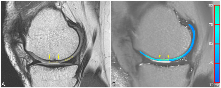
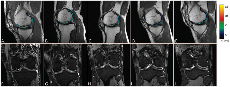
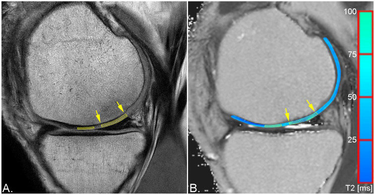
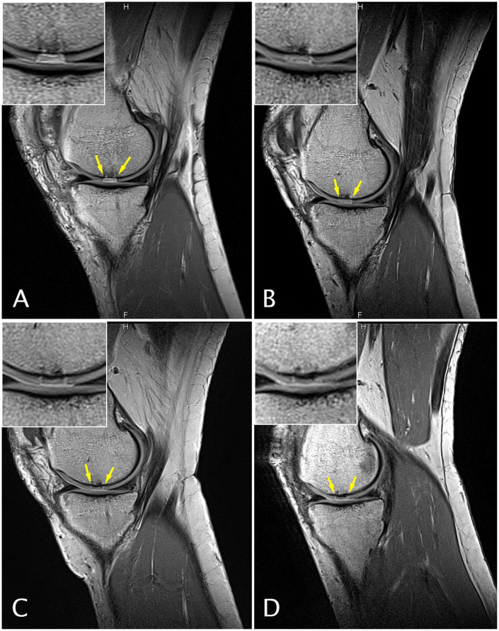
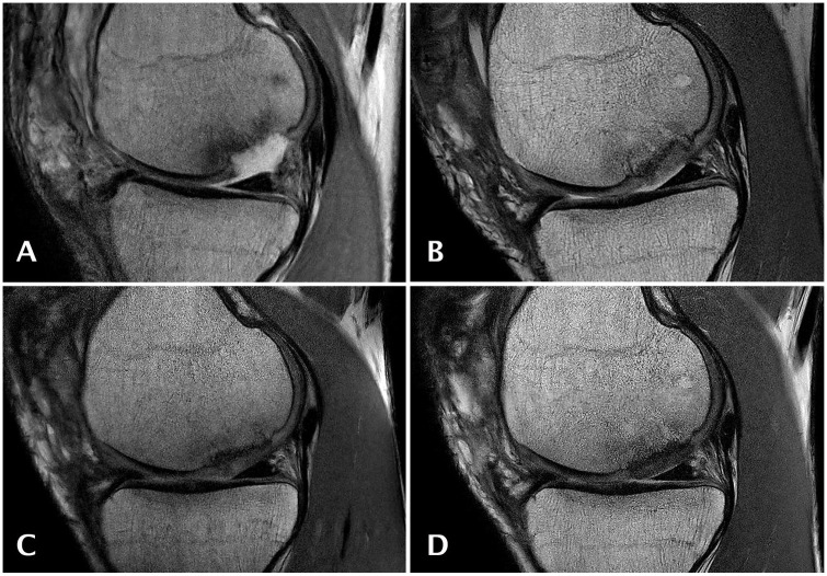
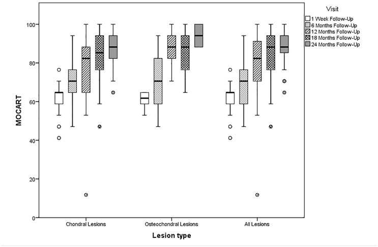
This study was designed as a prospective single-arm, open label, multicenter study. Morphological magnetic resonance imaging (MRI) for MOCART assessment and T mapping was performed 1 week and 6, 12, 18, and 24 months after GelrinC implantation. Evaluation of T mapping was based on the assessment of global T indices (T of the repair tissue [RT] divided by T of healthy reference cartilage) and zonal variation.
Fifty-six (20 female) patients were prospectively enrolled. The mean MOCART score significantly increased from baseline to the 24-month follow-up with 88.8 (95% CI, 85.8-91.9; < 0.001) for all lesions combined as well as 86.8 (95% CI, 83.0-90.6) for chondral lesions and 94.1 (95% CI, 68.55-100) for osteochondral lesions. Furthermore, based on T mapping, significant zonal variation of the RT was observed at 24 months ( = 0.039), which did not differ significantly from healthy reference cartilage ( = 0.6).
Increasing MOCART scores were observed throughout the follow-up period, indicative of maturation of the cartilage repair. Significant zonal variation of the RT at 24 months might indicate the transformation into hyaline cartilage-like RT. Slightly differing morphological outcome between chondral and osteochondral lesions, but similar global and zonal T indices at 24 months, support the potential of GelrinC as a treatment option for both lesion types.
使用形态学(软骨修复组织磁共振观察法[MOCART])和定量(T映射)磁共振成像(MRI)前瞻性评估GelrinC治疗股骨软骨和骨软骨损伤的疗效。
本研究设计为前瞻性单臂、开放标签、多中心研究。在植入GelrinC后1周以及6、12、18和24个月进行用于MOCART评估的形态学磁共振成像(MRI)和T映射。T映射评估基于整体T指数(修复组织[RT]的T除以健康对照软骨的T)和区域变化。
前瞻性纳入了56例(20例女性)患者。所有损伤合并后的平均MOCART评分从基线到24个月随访时显著增加,为88.8(95%置信区间,85.8 - 91.9;P < 0.001),软骨损伤为86.8(95%置信区间,83.0 - 90.6),骨软骨损伤为94.1(95%置信区间,68.55 - 100)。此外,基于T映射,在24个月时观察到RT有显著的区域变化(P = 0.039),与健康对照软骨无显著差异(P = 0.6)。
在整个随访期间观察到MOCART评分增加,表明软骨修复成熟。24个月时RT有显著的区域变化可能表明向透明软骨样RT转变。软骨损伤和骨软骨损伤之间形态学结果略有不同,但24个月时整体和区域T指数相似,支持GelrinC作为两种损伤类型治疗选择的潜力。