Center for Neurobiology Research, Fralin Biomedical Research Institute at Virginia Tech Carilion, Roanoke, VA, USA.
Graduate Program in Translational Biology, Medicine, and Health, Virginia Tech, Blacksburg, VA, USA.
J Neurochem. 2021 Nov;159(3):479-497. doi: 10.1111/jnc.15101. Epub 2020 Jun 24.
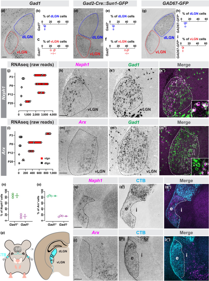
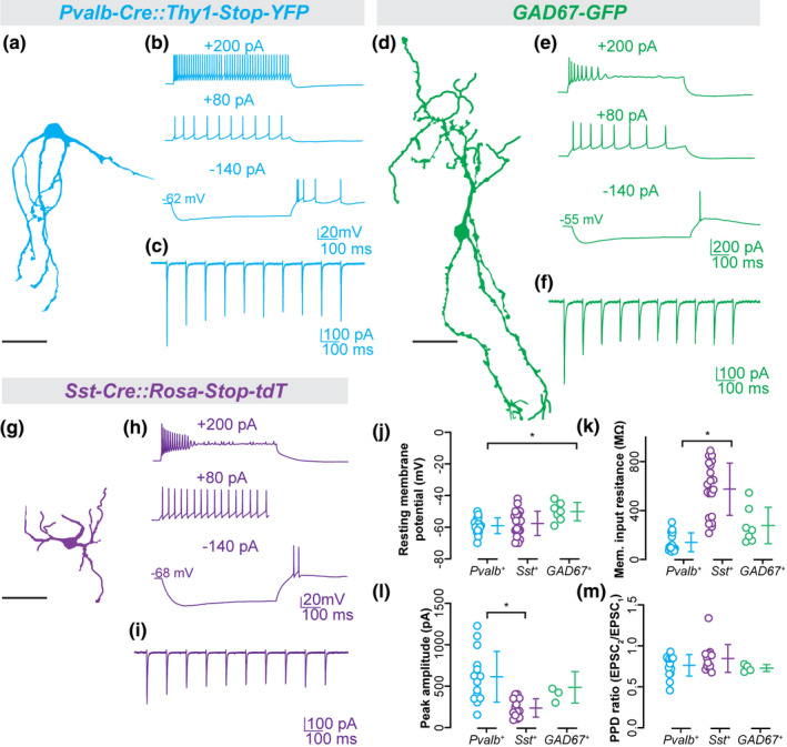
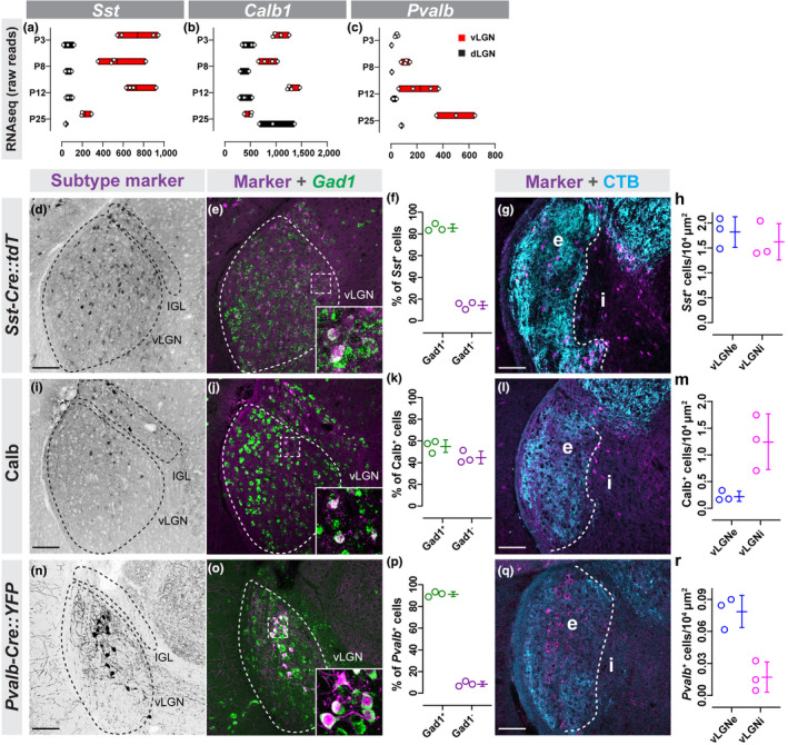
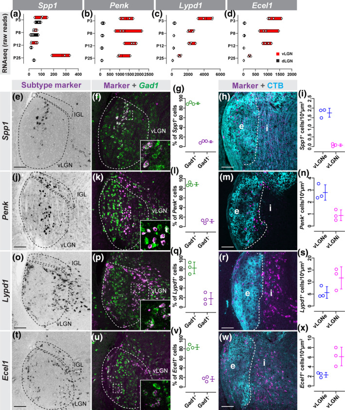
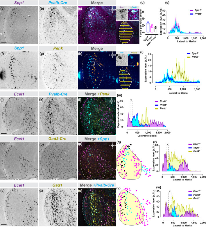
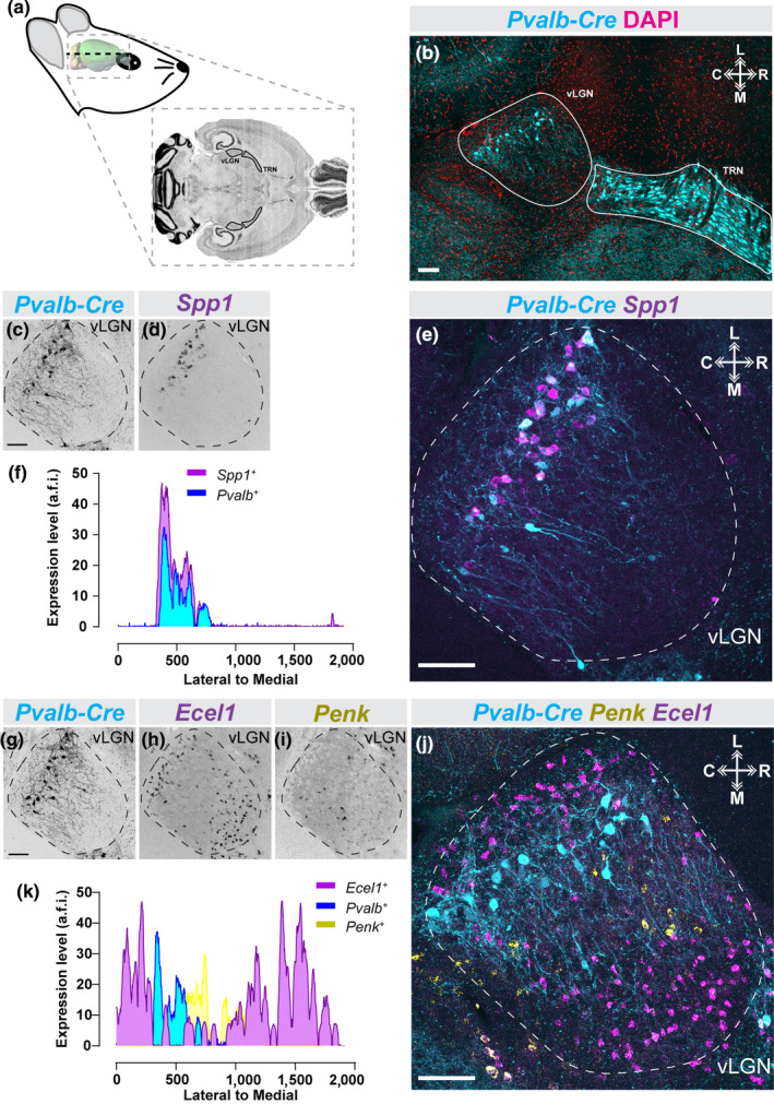
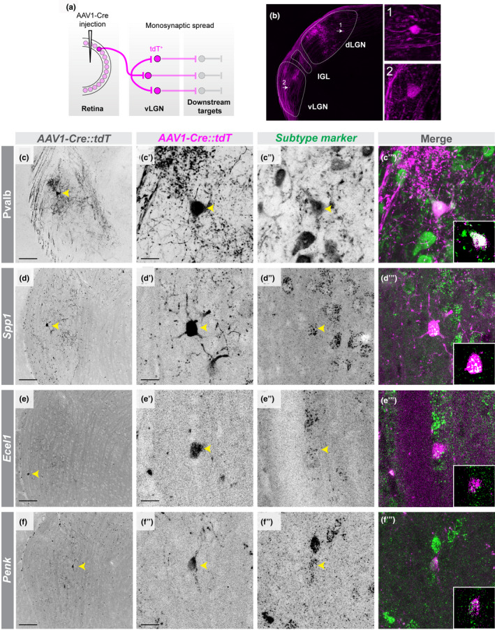
In the visual system, retinal axons convey visual information from the outside world to dozens of distinct retinorecipient brain regions and organize that information at several levels, including either at the level of retinal afferents, cytoarchitecture of intrinsic retinorecipient neurons, or a combination of the two. Two major retinorecipient nuclei which are densely innervated by retinal axons are the dorsal lateral geniculate nucleus, which is important for classical image-forming vision, and ventral LGN (vLGN), which is associated with non-image-forming vision. The neurochemistry, cytoarchitecture, and retinothalamic connectivity in vLGN remain unresolved, raising fundamental questions of how it receives and processes visual information. To shed light on these important questions, used in situ hybridization, immunohistochemistry, and genetic reporter lines to identify and characterize novel neuronal cell types in mouse vLGN. Not only were a high percentage of these cells GABAergic, we discovered transcriptomically distinct GABAergic cell types reside in the two major laminae of vLGN, the retinorecipient, external vLGN (vLGNe) and the non-retinorecipient, internal vLGN (vLGNi). Furthermore, within vLGNe, we identified transcriptionally distinct subtypes of GABAergic cells that are distributed into four adjacent sublaminae. Using trans-synaptic viral tracing and in vitro electrophysiology, we found cells in each these vLGNe sublaminae receive monosynaptic inputs from retina. These results not only identify novel subtypes of GABAergic cells in vLGN, they suggest the subtype-specific laminar distribution of retinorecipient cells in vLGNe may be important for receiving, processing, and transmitting light-derived signals in parallel channels of the subcortical visual system.
在视觉系统中,视网膜轴突将来自外部世界的视觉信息传递到数十个不同的视网膜接受脑区,并在多个层次上对信息进行组织,包括在视网膜传入纤维、内在视网膜接受神经元的细胞结构或两者的组合水平上进行组织。两个主要的视网膜接受核是背外侧膝状体核(dorsal lateral geniculate nucleus,dLGN),它对经典的成像视觉很重要,以及腹外侧膝状体核(ventral LGN,vLGN),它与非成像视觉有关。vLGN 的神经化学、细胞结构和视网膜丘脑连接仍然没有得到解决,这就提出了一个基本问题,即它如何接收和处理视觉信息。为了解决这些重要问题,我们使用原位杂交、免疫组织化学和遗传报告基因系来鉴定和描述小鼠 vLGN 中的新型神经元细胞类型。不仅这些细胞中有很大比例是 GABA 能的,我们还发现,在 vLGN 的两个主要层,即视网膜接受的外部 vLGN(vLGNe)和非视网膜接受的内部 vLGN(vLGNi)中,存在转录上不同的 GABA 能细胞类型。此外,在 vLGNe 中,我们鉴定了转录上不同的 GABA 能细胞亚型,它们分布在四个相邻的亚层中。通过跨突触病毒追踪和体外电生理学,我们发现每个 vLGNe 亚层中的细胞都从视网膜接收单突触输入。这些结果不仅鉴定了 vLGN 中新型的 GABA 能细胞亚型,还表明 vLGNe 中视网膜接受细胞的亚型特异性层分布可能对在亚皮质视觉系统的并行通道中接收、处理和传递光衍生信号很重要。