Media Lab, Massachusetts Institute of Technology (MIT), Cambridge, Massachusetts.
McGovern Institute, MIT, Cambridge, Massachusetts.
Curr Protoc Neurosci. 2020 Jun;92(1):e96. doi: 10.1002/cpns.96.
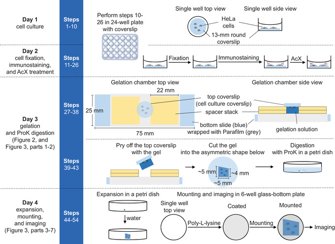
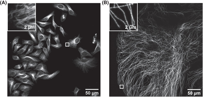
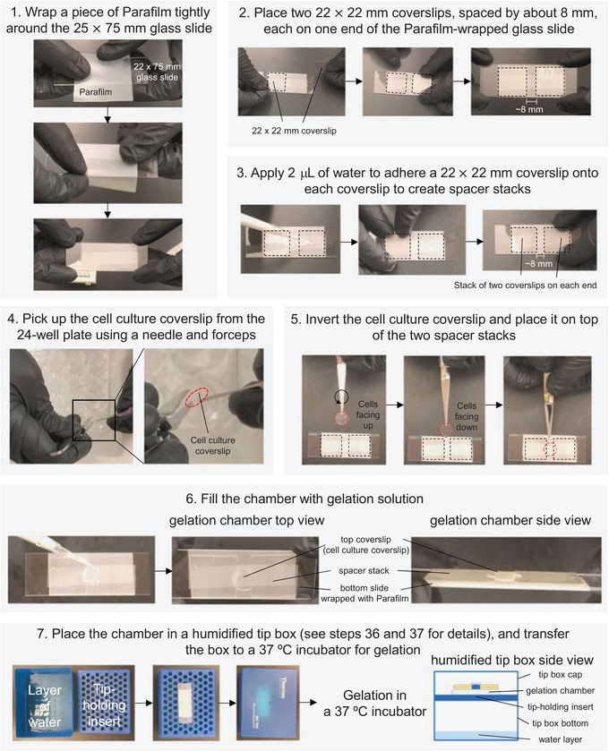
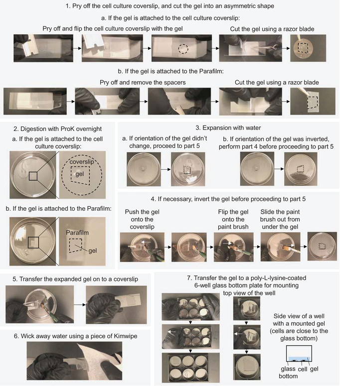
Expansion microscopy (ExM) is a technique that physically expands preserved cells and tissues before microscope imaging, so that conventional diffraction-limited microscopes can perform nanoscale-resolution imaging. In ExM, biomolecules or their markers are linked to a dense, swellable gel network synthesized throughout a specimen. Mechanical homogenization of the sample (e.g., by protease digestion) and the addition of water enable isotropic swelling of the gel, so that the relative positions of biomolecules are preserved. We previously presented ExM protocols for analyzing proteins and RNAs in cells and tissues. Here we describe a cookbook-style ExM protocol for expanding cultured HeLa cells with immunostained microtubules, aimed to help newcomers familiarize themselves with the experimental setups and skills required to successfully perform ExM. Our aim is to help beginners, or students in a wet-lab classroom setting, learn all the key steps of ExM. © 2020 The Authors.
扩展显微镜技术(ExM)是一种在显微镜成像之前对保存的细胞和组织进行物理扩展的技术,使得传统的衍射极限显微镜能够进行纳米分辨率成像。在 ExM 中,生物分子或其标记物与整个标本中合成的密集、可溶胀的凝胶网络连接。通过机械均化样品(例如,通过蛋白酶消化)和添加水,使凝胶能够各向同性地溶胀,从而保留生物分子的相对位置。我们之前提出了用于分析细胞和组织中的蛋白质和 RNA 的 ExM 方案。在这里,我们描述了一种用于扩展免疫染色微管的培养 HeLa 细胞的扩展显微镜技术(ExM)方案,旨在帮助初学者熟悉成功执行 ExM 所需的实验设置和技能。我们的目标是帮助初学者或在湿实验室课堂环境中的学生学习 ExM 的所有关键步骤。