• 文献检索
  • 文档翻译
  • 深度研究
  • 学术资讯
  • Suppr Zotero 插件Zotero 插件
  • 邀请有礼
  • 套餐&价格
  • 历史记录
应用&插件
Suppr Zotero 插件Zotero 插件浏览器插件Mac 客户端Windows 客户端微信小程序
定价
高级版会员购买积分包购买API积分包
服务
文献检索文档翻译深度研究API 文档MCP 服务
关于我们
关于 Suppr公司介绍联系我们用户协议隐私条款
关注我们

Suppr 超能文献

核心技术专利:CN118964589B侵权必究
粤ICP备2023148730 号-1Suppr @ 2026

文献检索

告别复杂PubMed语法,用中文像聊天一样搜索,搜遍4000万医学文献。AI智能推荐,让科研检索更轻松。

立即免费搜索

文件翻译

保留排版,准确专业,支持PDF/Word/PPT等文件格式,支持 12+语言互译。

免费翻译文档

深度研究

AI帮你快速写综述,25分钟生成高质量综述,智能提取关键信息,辅助科研写作。

立即免费体验

X10 扩展显微镜可在常规显微镜上实现 25nm 的分辨率。

X10 expansion microscopy enables 25-nm resolution on conventional microscopes.

机构信息

Institute for Neuro- and Sensory Physiology, Center for Biostructural Imaging of Neurodegeneration, Cluster of Excellence Nanoscale Microscopy and Molecular Physiology of the Brain, University Medical Center Göttingen, Göttingen, Germany

International Max Planck Research School for Molecular Biology, Göttingen, Germany.

出版信息

EMBO Rep. 2018 Sep;19(9). doi: 10.15252/embr.201845836. Epub 2018 Jul 9.

DOI:10.15252/embr.201845836
PMID:29987134
原文链接:https://pmc.ncbi.nlm.nih.gov/articles/PMC6123658/
Abstract

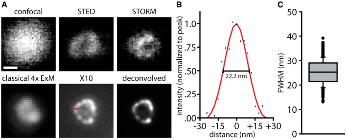
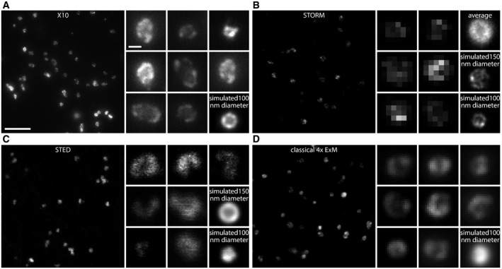
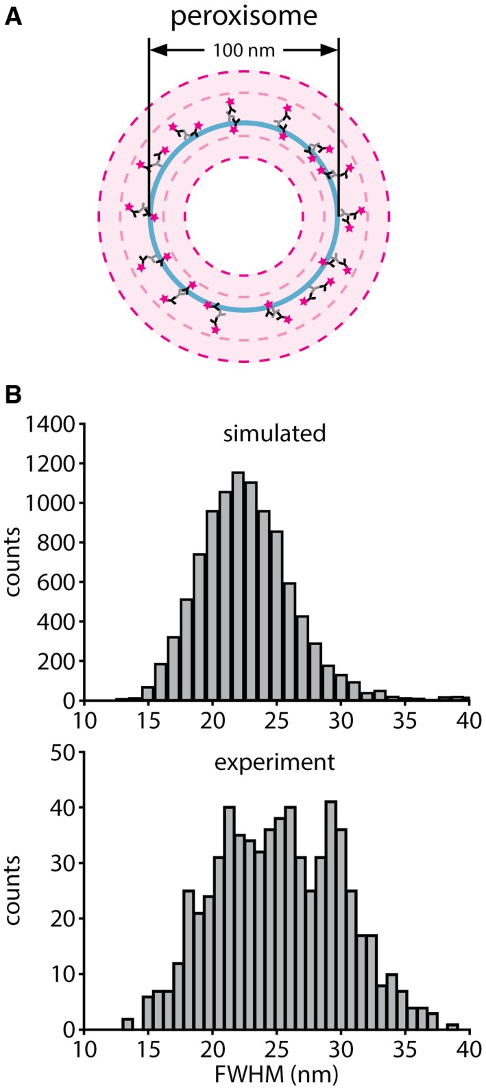
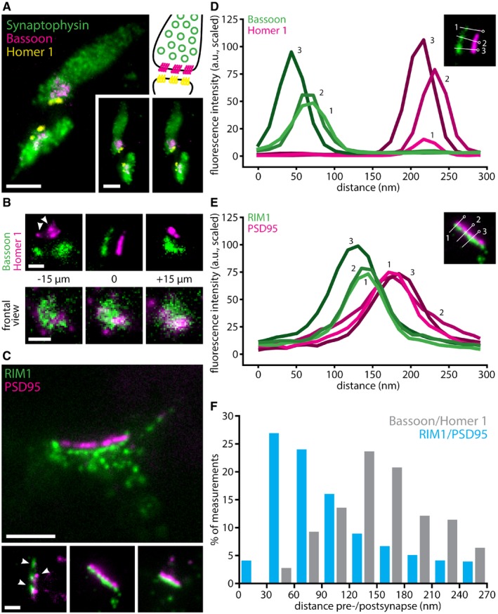
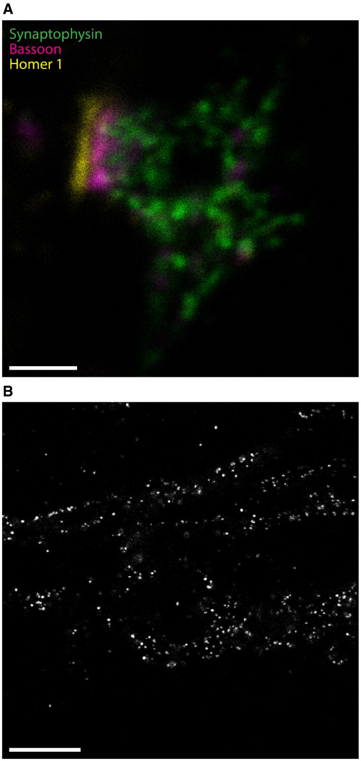
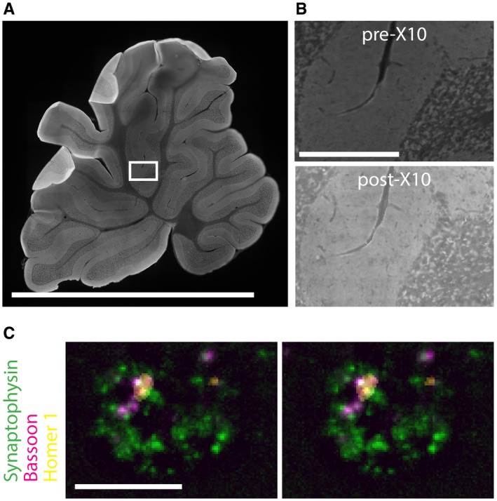
Expansion microscopy is a recently introduced imaging technique that achieves super-resolution through physically expanding the specimen by ~4×, after embedding into a swellable gel. The resolution attained is, correspondingly, approximately fourfold better than the diffraction limit, or ~70 nm. This is a major improvement over conventional microscopy, but still lags behind modern STED or STORM setups, whose resolution can reach 20-30 nm. We addressed this issue here by introducing an improved gel recipe that enables an expansion factor of ~10× in each dimension, which corresponds to an expansion of the sample volume by more than 1,000-fold. Our protocol, which we termed X10 microscopy, achieves a resolution of 25-30 nm on conventional epifluorescence microscopes. X10 provides multi-color images similar or even superior to those produced with more challenging methods, such as STED, STORM, and iterative expansion microscopy (iExM). X10 is therefore the cheapest and easiest option for high-quality super-resolution imaging currently available. X10 should be usable in any laboratory, irrespective of the machinery owned or of the technical knowledge.

摘要

扩展显微镜是一种最近引入的成像技术,通过在嵌入可溶胀凝胶后将样品物理膨胀约 4 倍来实现超分辨率。相应地,获得的分辨率比衍射极限大约高出四倍,约为 70nm。这是对传统显微镜的重大改进,但仍落后于现代 STED 或 STORM 设备,其分辨率可达到 20-30nm。在这里,我们通过引入一种改进的凝胶配方来解决这个问题,该配方在每个维度上都能实现约 10 倍的扩展因子,这对应于样品体积的扩展超过 1000 倍。我们的方法称为 X10 显微镜,在传统的荧光显微镜上实现了 25-30nm 的分辨率。X10 提供的多色图像与 STED、STORM 和迭代扩展显微镜(iExM)等更具挑战性的方法产生的图像相似,甚至更好。因此,X10 是目前可用的最高质量超分辨率成像的最便宜、最简单的选择。X10 应该可以在任何实验室使用,无论其拥有的设备或技术知识如何。

https://cdn.ncbi.nlm.nih.gov/pmc/blobs/f82c/6123658/097b757b46ef/EMBR-19-e45836-g011.jpg
https://cdn.ncbi.nlm.nih.gov/pmc/blobs/f82c/6123658/85c5c947e162/EMBR-19-e45836-g002.jpg
https://cdn.ncbi.nlm.nih.gov/pmc/blobs/f82c/6123658/5c16658e01bf/EMBR-19-e45836-g004.jpg
https://cdn.ncbi.nlm.nih.gov/pmc/blobs/f82c/6123658/260d6ba80495/EMBR-19-e45836-g003.jpg
https://cdn.ncbi.nlm.nih.gov/pmc/blobs/f82c/6123658/c25891ae647e/EMBR-19-e45836-g006.jpg
https://cdn.ncbi.nlm.nih.gov/pmc/blobs/f82c/6123658/435d2c382b6b/EMBR-19-e45836-g005.jpg
https://cdn.ncbi.nlm.nih.gov/pmc/blobs/f82c/6123658/bbea520e5c04/EMBR-19-e45836-g007.jpg
https://cdn.ncbi.nlm.nih.gov/pmc/blobs/f82c/6123658/535394d69bd1/EMBR-19-e45836-g008.jpg
https://cdn.ncbi.nlm.nih.gov/pmc/blobs/f82c/6123658/52b18b65e1d9/EMBR-19-e45836-g009.jpg
https://cdn.ncbi.nlm.nih.gov/pmc/blobs/f82c/6123658/578762f3f222/EMBR-19-e45836-g010.jpg
https://cdn.ncbi.nlm.nih.gov/pmc/blobs/f82c/6123658/097b757b46ef/EMBR-19-e45836-g011.jpg

相似文献

1
X10 expansion microscopy enables 25-nm resolution on conventional microscopes.X10 扩展显微镜可在常规显微镜上实现 25nm 的分辨率。
EMBO Rep. 2018 Sep;19(9). doi: 10.15252/embr.201845836. Epub 2018 Jul 9.
2
Simple multi-color super-resolution by X10 microscopy.X10 显微镜实现简单的多色超分辨率。
Methods Cell Biol. 2021;161:33-56. doi: 10.1016/bs.mcb.2020.04.016. Epub 2020 May 27.
3
A practical guide to optimization in X10 expansion microscopy.X10 扩展显微镜优化实用指南。
Nat Protoc. 2019 Mar;14(3):832-863. doi: 10.1038/s41596-018-0117-3.
4
Amplified Expansion Stimulated Emission Depletion Microscopy.放大扩展受激发射损耗显微镜。
Chembiochem. 2019 May 15;20(10):1260-1265. doi: 10.1002/cbic.201800775. Epub 2019 Apr 8.
5
Super-resolution STED microscopy in live brain tissue.活脑组织中的超高分辨率 STED 显微镜。
Neurobiol Dis. 2021 Aug;156:105420. doi: 10.1016/j.nbd.2021.105420. Epub 2021 Jun 5.
6
Heat denaturation enables multicolor X10-STED microscopy.热变性使多色 X10-STED 显微镜成为可能。
Sci Rep. 2023 Apr 1;13(1):5366. doi: 10.1038/s41598-023-32524-5.
7
ExCel: Super-Resolution Imaging of C. elegans with Expansion Microscopy.ExCel:利用扩展显微镜对秀丽隐杆线虫进行超分辨率成像。
Methods Mol Biol. 2022;2468:141-203. doi: 10.1007/978-1-0716-2181-3_9.
8
Challenges of Using Expansion Microscopy for Super-resolved Imaging of Cellular Organelles.使用扩展显微镜进行细胞细胞器超分辨成像的挑战。
Chembiochem. 2021 Feb 15;22(4):686-693. doi: 10.1002/cbic.202000571. Epub 2020 Nov 11.
9
Iterative expansion microscopy.迭代扩展显微镜技术。
Nat Methods. 2017 Jun;14(6):593-599. doi: 10.1038/nmeth.4261. Epub 2017 Apr 17.
10
Fast live-cell conventional fluorophore nanoscopy with ImageJ through super-resolution radial fluctuations.基于超分辨率径向波动的 ImageJ 快速活细胞常规荧光纳米显微镜
Nat Commun. 2016 Aug 12;7:12471. doi: 10.1038/ncomms12471.

引用本文的文献

1
Functional synapses between neurons and small cell lung cancer.神经元与小细胞肺癌之间的功能性突触。
Nature. 2025 Sep 10. doi: 10.1038/s41586-025-09434-9.
2
Expansion and fluctuations-enhanced microscopy for nanoscale molecular profiling of cells and tissues.用于细胞和组织纳米级分子分析的扩展与波动增强显微镜技术。
Nat Protoc. 2025 Jul 2. doi: 10.1038/s41596-025-01178-0.
3
Expansion-assisted selective plane illumination microscopy for nanoscale imaging of centimeter-scale tissues.用于厘米级组织纳米尺度成像的扩展辅助选择性平面照明显微术。

本文引用的文献

1
Hybrid Structured Illumination Expansion Microscopy Reveals Microbial Cytoskeleton Organization.杂交结构光照扩展显微镜揭示微生物细胞骨架组织。
ACS Nano. 2017 Dec 26;11(12):12677-12686. doi: 10.1021/acsnano.7b07200. Epub 2017 Nov 30.
2
Nanoscale imaging of clinical specimens using pathology-optimized expansion microscopy.使用病理学优化的扩张显微镜对临床标本进行纳米级成像。
Nat Biotechnol. 2017 Aug;35(8):757-764. doi: 10.1038/nbt.3892. Epub 2017 Jul 17.
3
Q&A: Expansion microscopy.问答:扩展显微镜技术
Elife. 2025 Jun 30;12:RP91979. doi: 10.7554/eLife.91979.
4
Endogenous SNAP-Tagging of Munc13‑1 for Monitoring Synapse Nanoarchitecture.用于监测突触纳米结构的Munc13-1内源性SNAP标记
JACS Au. 2025 May 23;5(6):2475-2490. doi: 10.1021/jacsau.4c00946. eCollection 2025 Jun 23.
5
A parallelly distributed microscope and software system for scalable high-throughput multispectral 3D imaging.一种用于可扩展高通量多光谱三维成像的并行分布式显微镜和软件系统。
bioRxiv. 2025 Jun 3:2025.05.31.657163. doi: 10.1101/2025.05.31.657163.
6
Light-microscopy-based connectomic reconstruction of mammalian brain tissue.基于光学显微镜的哺乳动物脑组织连接组重建
Nature. 2025 May 7. doi: 10.1038/s41586-025-08985-1.
7
Microtubules as a versatile reference standard for expansion microscopy.微管作为扩展显微镜术的通用参考标准
Commun Biol. 2025 Mar 26;8(1):499. doi: 10.1038/s42003-025-07967-3.
8
Super-resolution Analysis of Astrocyte Morphology Using Expansion Microscopy.使用扩展显微镜对星形胶质细胞形态进行超分辨率分析。
Methods Mol Biol. 2025;2896:165-179. doi: 10.1007/978-1-0716-4366-2_12.
9
BOOST: a robust ten-fold expansion method on hour-scale.BOOST:一种在小时尺度上的稳健的十倍扩增方法。
Nat Commun. 2025 Mar 2;16(1):2107. doi: 10.1038/s41467-025-57350-3.
10
Seq-Scope-eXpanded: Spatial Omics Beyond Optical Resolution.Seq-Scope扩展版:超越光学分辨率的空间组学
bioRxiv. 2025 Feb 8:2025.02.04.636355. doi: 10.1101/2025.02.04.636355.
BMC Biol. 2017 Jun 19;15(1):50. doi: 10.1186/s12915-017-0393-3.
4
Iterative expansion microscopy.迭代扩展显微镜技术。
Nat Methods. 2017 Jun;14(6):593-599. doi: 10.1038/nmeth.4261. Epub 2017 Apr 17.
5
Nanometer resolution imaging and tracking of fluorescent molecules with minimal photon fluxes.纳米分辨率荧光分子的成像和跟踪,使用最小的光子通量。
Science. 2017 Feb 10;355(6325):606-612. doi: 10.1126/science.aak9913. Epub 2016 Dec 22.
6
A trans-synaptic nanocolumn aligns neurotransmitter release to receptors.一个跨突触纳米柱将神经递质释放与受体对齐。
Nature. 2016 Aug 11;536(7615):210-4. doi: 10.1038/nature19058. Epub 2016 Jul 27.
7
Nanoscale imaging of RNA with expansion microscopy.利用扩张显微镜对RNA进行纳米级成像。
Nat Methods. 2016 Aug;13(8):679-84. doi: 10.1038/nmeth.3899. Epub 2016 Jul 4.
8
Protein-retention expansion microscopy of cells and tissues labeled using standard fluorescent proteins and antibodies.使用标准荧光蛋白和抗体标记的细胞和组织的蛋白质保留扩展显微镜技术。
Nat Biotechnol. 2016 Sep;34(9):987-92. doi: 10.1038/nbt.3625. Epub 2016 Jul 4.
9
Imaging cellular structures in super-resolution with SIM, STED and Localisation Microscopy: A practical comparison.利用结构照明显微镜(SIM)、受激发射损耗显微镜(STED)和定位显微镜进行超分辨率细胞结构成像:实际比较
Sci Rep. 2016 Jun 6;6:27290. doi: 10.1038/srep27290.
10
Expansion microscopy with conventional antibodies and fluorescent proteins.使用传统抗体和荧光蛋白的扩展显微镜技术。
Nat Methods. 2016 Jun;13(6):485-8. doi: 10.1038/nmeth.3833. Epub 2016 Apr 11.