• 文献检索
  • 文档翻译
  • 深度研究
  • 学术资讯
  • Suppr Zotero 插件Zotero 插件
  • 邀请有礼
  • 套餐&价格
  • 历史记录
应用&插件
Suppr Zotero 插件Zotero 插件浏览器插件Mac 客户端Windows 客户端微信小程序
定价
高级版会员购买积分包购买API积分包
服务
文献检索文档翻译深度研究API 文档MCP 服务
关于我们
关于 Suppr公司介绍联系我们用户协议隐私条款
关注我们

Suppr 超能文献

核心技术专利:CN118964589B侵权必究
粤ICP备2023148730 号-1Suppr @ 2026

文献检索

告别复杂PubMed语法,用中文像聊天一样搜索,搜遍4000万医学文献。AI智能推荐,让科研检索更轻松。

立即免费搜索

文件翻译

保留排版,准确专业,支持PDF/Word/PPT等文件格式,支持 12+语言互译。

免费翻译文档

深度研究

AI帮你快速写综述,25分钟生成高质量综述,智能提取关键信息,辅助科研写作。

立即免费体验

肥厚型心肌病的T2加权心脏磁共振成像与心肌生物标志物

T2-weighted cardiac magnetic resonance image and myocardial biomarker in hypertrophic cardiomyopathy.

作者信息

Chen Shi, Huang Liwei, Zhang Qing, Wang Jie, Chen Yucheng

机构信息

Department of Cardiology, West China Hospital, Sichuan University.

Department of Cardiovascular Ultrasound and Noninvasive Cardiology, Sichuan Academy of Medical Sciences and Sichuan People's Hospital, Chengdu, Sichuan, China.

出版信息

Medicine (Baltimore). 2020 Jun 5;99(23):e20134. doi: 10.1097/MD.0000000000020134.

DOI:10.1097/MD.0000000000020134
PMID:32501969
原文链接:https://pmc.ncbi.nlm.nih.gov/articles/PMC7306317/
Abstract

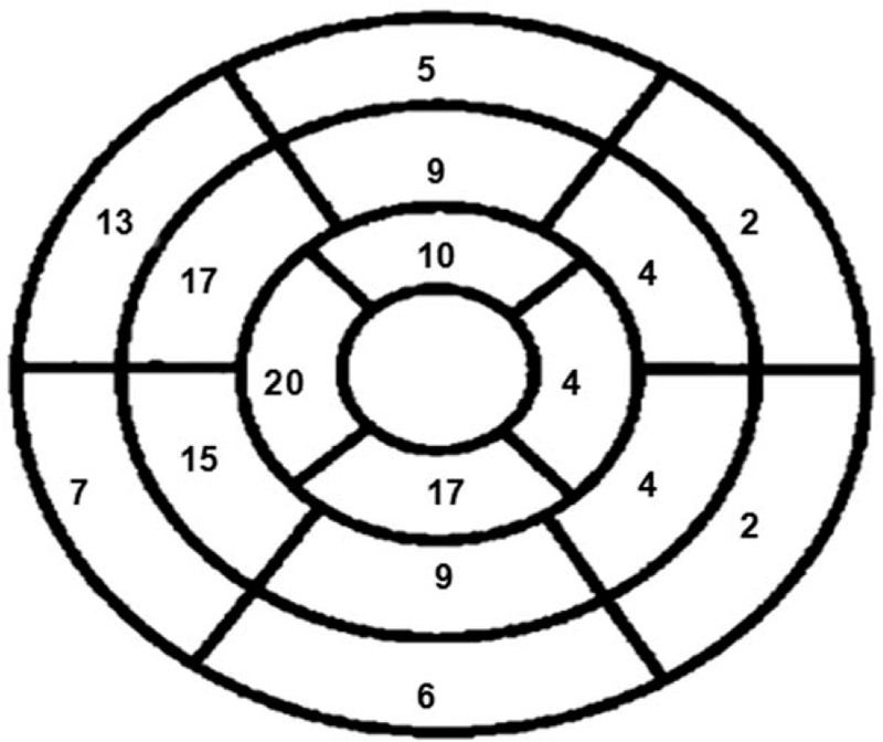
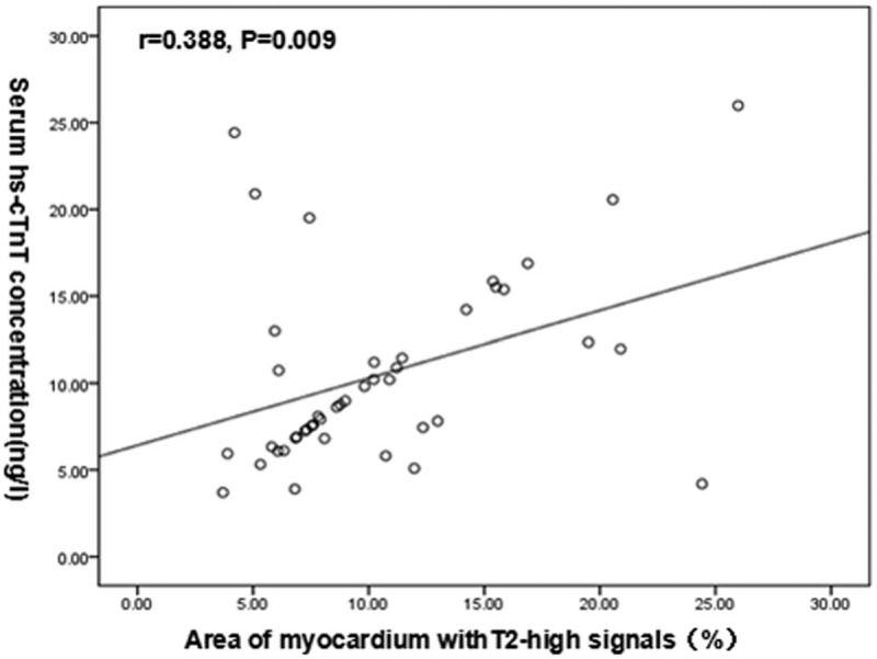
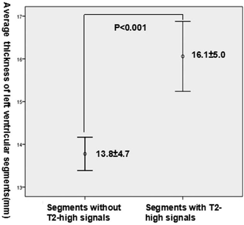
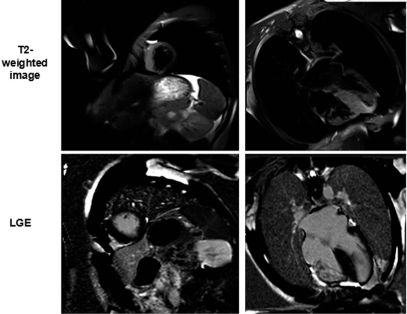
The phenomenon of high signal intensity on T2-weighted imaging of cardiac magnetic resonance in hypertrophic cardiomyopathy (HCM) has been previously studied. However, the underlying histopathologic mechanism remains unclear. Elevated cardiac troponin can be detected in some HCM patients. A reasonable hypothesis is that high myocardial T2 signal is a potential marker of myocardial injury in HCM. We sought to investigate the association between cardiac troponin and the extent of high T2 signals in HCM patients.Forty-four HCM patients underwent 3.0T cardiac magnetic resonance scanning. On T2-weighted images, the number of segments with high-signal intensity (myocardium-to-skeletal muscle signal intensity ratio >2) and the percentage of high-signal area (>2 standard deviation above the remote tissue) were measured in 16 myocardial segments along the LV mid-myocardial circumference on 3 short-axis images. The level of high-sensitivity cardiac troponin T (hs-cTnT) was also assessed.Myocardial high T2 signals were identified in 33 (75%) patients and 144 (20.5%) segments. Elevated hs-cTnT was observed in 28 (63.6%) patients. The Cochran-Armitage test showed a statistically significant trend of increasing levels of hs-cTnT with elevated number of segments with myocardial high T2 signal (P = .002). Further, the percentage of myocardium with high T2 signal was significantly associated with the hs-cTnT level (Pearson correlation: r = 0.388, P = .009).Myocardium with high T2 signals was very common in patients with HCM.Its extent is related with the level of plasma hs-cTnT.

摘要

肥厚型心肌病(HCM)患者心脏磁共振T2加权成像上高信号强度现象此前已有研究。然而,其潜在的组织病理学机制仍不清楚。部分HCM患者可检测到心肌肌钙蛋白升高。一个合理的假设是,心肌T2高信号是HCM中心肌损伤的潜在标志物。我们试图研究HCM患者中心肌肌钙蛋白与T2高信号范围之间的关联。

44例HCM患者接受了3.0T心脏磁共振扫描。在T2加权图像上,于3幅短轴图像上沿左心室心肌中层圆周的16个心肌节段测量高信号强度节段数量(心肌与骨骼肌信号强度比>2)以及高信号面积百分比(高于远隔组织2个标准差以上)。同时评估高敏心肌肌钙蛋白T(hs-cTnT)水平。

33例(75%)患者及144个(20.5%)节段发现心肌T2高信号。28例(63.6%)患者hs-cTnT升高。 Cochr an-Armitage检验显示,随着心肌T2高信号节段数量增加,hs-cTnT水平呈统计学显著升高趋势(P = 0.002)。此外,T2高信号心肌百分比与hs-cTnT水平显著相关(Pearson相关系数:r = 0.388,P = 0.009)。

HCM患者中T2高信号心肌很常见。其范围与血浆hs-cTnT水平相关。

https://cdn.ncbi.nlm.nih.gov/pmc/blobs/d23c/7306317/98f855c1c617/medi-99-e20134-g006.jpg
https://cdn.ncbi.nlm.nih.gov/pmc/blobs/d23c/7306317/5fcb91c54a31/medi-99-e20134-g003.jpg
https://cdn.ncbi.nlm.nih.gov/pmc/blobs/d23c/7306317/a768dd87fce6/medi-99-e20134-g004.jpg
https://cdn.ncbi.nlm.nih.gov/pmc/blobs/d23c/7306317/134956c63e30/medi-99-e20134-g005.jpg
https://cdn.ncbi.nlm.nih.gov/pmc/blobs/d23c/7306317/98f855c1c617/medi-99-e20134-g006.jpg

相似文献

1
T2-weighted cardiac magnetic resonance image and myocardial biomarker in hypertrophic cardiomyopathy.肥厚型心肌病的T2加权心脏磁共振成像与心肌生物标志物
Medicine (Baltimore). 2020 Jun 5;99(23):e20134. doi: 10.1097/MD.0000000000020134.
2
High T2-weighted signal intensity is associated with elevated troponin T in hypertrophic cardiomyopathy.在肥厚型心肌病中,高T2加权信号强度与肌钙蛋白T升高有关。
Heart. 2017 Feb 15;103(4):293-299. doi: 10.1136/heartjnl-2016-309900. Epub 2016 Aug 9.
3
Significance of high-sensitivity cardiac troponin T in hypertrophic cardiomyopathy.高敏心肌肌钙蛋白 T 在肥厚型心肌病中的意义。
J Am Coll Cardiol. 2013 Oct 1;62(14):1252-1259. doi: 10.1016/j.jacc.2013.03.055. Epub 2013 Apr 23.
4
Diffusion-weighted imaging in hypertrophic cardiomyopathy: association with high T2-weighted signal intensity in addition to late gadolinium enhancement.肥厚型心肌病的扩散加权成像:除延迟钆增强外,还与高T2加权信号强度相关。
Int J Cardiovasc Imaging. 2020 Nov;36(11):2229-2238. doi: 10.1007/s10554-020-01933-2. Epub 2020 Jul 14.
5
Circulating biomarkers of hypertrophy and fibrosis in patients with hypertrophic cardiomyopathy assessed by cardiac magnetic resonance.通过心脏磁共振评估肥厚型心肌病患者中肥大和纤维化的循环生物标志物。
Biomarkers. 2018 Nov;23(7):676-682. doi: 10.1080/1354750X.2018.1474261. Epub 2018 May 23.
6
Prevalence and determinants of elevated high-sensitivity cardiac troponin T in hypertrophic cardiomyopathy.肥厚型心肌病中心脏肌钙蛋白 T 高敏升高的患病率及其决定因素。
J Cardiol. 2014 Feb;63(2):140-4. doi: 10.1016/j.jjcc.2013.07.008. Epub 2013 Sep 4.
7
Exercise and myocardial injury in hypertrophic cardiomyopathy.肥厚型心肌病中的运动与心肌损伤
Heart. 2020 Aug;106(15):1169-1175. doi: 10.1136/heartjnl-2019-315818. Epub 2020 Jan 30.
8
MRI native T1 and T2 mapping of myocardial segments in hypertrophic cardiomyopathy: tissue remodeling manifested prior to structure changes.MRI 心肌节段 native T1 和 T2 mapping 在肥厚型心肌病中的应用:结构改变前的组织重构表现。
Br J Radiol. 2019 Dec;92(1104):20190634. doi: 10.1259/bjr.20190634. Epub 2019 Oct 16.
9
Elevated high-sensitivity troponin T levels are associated with adverse cardiac remodelling and myocardial fibrosis in hypertrophic cardiomyopathy.高敏肌钙蛋白T水平升高与肥厚型心肌病患者心脏不良重塑及心肌纤维化相关。
Swiss Med Wkly. 2016 Mar 21;146:w14285. doi: 10.4414/smw.2016.14285. eCollection 2016.
10
Distribution and Clinical Significance of High Signal Intensity of the Myocardium on T2-Weighted Images in 2 Phenotypes of Hypertrophic Cardiomyopathy.肥厚型心肌病两种表型中心肌在T2加权图像上高信号强度的分布及临床意义
J Comput Assist Tomogr. 2015 Nov-Dec;39(6):951-5. doi: 10.1097/RCT.0000000000000296.

引用本文的文献

1
Novel Magnetic Resonance Imaging Tools for Hypertrophic Cardiomyopathy Risk Stratification.用于肥厚型心肌病风险分层的新型磁共振成像工具
Life (Basel). 2024 Jan 30;14(2):200. doi: 10.3390/life14020200.
2
Sarcoid heart disease and imaging.结节病性心脏病与影像学
Heart Rhythm O2. 2023 Nov 19;5(1):50-59. doi: 10.1016/j.hroo.2023.11.012. eCollection 2024 Jan.
3
Cardiac Multimodality Imaging in Hypertrophic Cardiomyopathy: What to Look for and When to Image.肥厚型心肌病的心脏多模态影像学:需要观察什么以及何时进行成像。

本文引用的文献

1
Cardiac troponin I is associated with non-sustained ventricular tachycardia in patients with hypertrophic obstructive cardiomyopathy.心肌肌钙蛋白I与肥厚性梗阻性心肌病患者的非持续性室性心动过速相关。
Heart Vessels. 2020 Jun;35(6):876-885. doi: 10.1007/s00380-019-01549-0. Epub 2020 Jan 6.
2
The role of Cardiac Magnetic Resonance (CMR) in the diagnosis of cardiomyopathy: A systematic review.心脏磁共振成像(CMR)在心肌病诊断中的作用:一项系统综述。
Malawi Med J. 2019 Sep;31(3):241-245. doi: 10.4314/mmj.v31i3.14.
3
The prognostic value of biventricular long axis strain using standard cardiovascular magnetic resonance imaging in patients with hypertrophic cardiomyopathy.
Curr Cardiol Rev. 2023;19(5):1-18. doi: 10.2174/1573403X19666230316103117.
4
Left ventricular strain patterns and their relationships with cardiac biomarkers in hypertrophic cardiomyopathy patients with preserved left ventricular ejection fraction.左心室射血分数保留的肥厚型心肌病患者的左心室应变模式及其与心脏生物标志物的关系。
Front Cardiovasc Med. 2022 Oct 4;9:963110. doi: 10.3389/fcvm.2022.963110. eCollection 2022.
应用标准心血管磁共振成像评估肥厚型心肌病患者的双心室长轴应变的预后价值。
Int J Cardiol. 2019 Nov 1;294:43-49. doi: 10.1016/j.ijcard.2019.08.010. Epub 2019 Aug 3.
4
High Signal Intensity on T2-Weighted Cardiovascular Magnetic Resonance Imaging Predicts Life-Threatening Arrhythmic Events in Hypertrophic Cardiomyopathy Patients.T2 加权心血管磁共振成像上的高信号强度可预测肥厚型心肌病患者的危及生命的心律失常事件。
Circ J. 2018 Mar 23;82(4):1062-1069. doi: 10.1253/circj.CJ-17-1235. Epub 2018 Feb 21.
5
Elevated Level of Troponin but Not N-Terminal Probrain Natriuretic Peptide Is Associated with Increased Risk of Sudden Cardiac Death in Hypertrophic Cardiomyopathy Calculated According to the ESC Guidelines 2014.根据 2014 年 ESC 指南,肌钙蛋白水平升高而非 N 末端脑利钠肽前体与肥厚型心肌病患者发生心源性猝死风险增加相关。
Dis Markers. 2017;2017:9417908. doi: 10.1155/2017/9417908. Epub 2017 Nov 20.
6
Phenotypic diversity identified by cardiac magnetic resonance in a large hypertrophic cardiomyopathy family with a single MYH7 mutation.通过心脏磁共振在一个具有单一 MYH7 突变的大型肥厚型心肌病家族中鉴定出的表型多样性。
Sci Rep. 2018 Jan 17;8(1):973. doi: 10.1038/s41598-018-19372-4.
7
CMR assessment and clinical outcomes of hypertrophic cardiomyopathy with or without ventricular remodeling in the end-stage phase.终末期肥厚型心肌病伴或不伴心室重构的心脏磁共振成像评估及临床结局
Int J Cardiovasc Imaging. 2018 Apr;34(4):597-605. doi: 10.1007/s10554-017-1263-3. Epub 2017 Oct 25.
8
High T2-weighted signal intensity for risk prediction of sudden cardiac death in hypertrophic cardiomyopathy.肥厚型心肌病中用于预测心源性猝死风险的高T2加权信号强度
Int J Cardiovasc Imaging. 2018 Jan;34(1):113-120. doi: 10.1007/s10554-017-1252-6. Epub 2017 Oct 23.
9
Myocardial Edema on T2-Weighted MRI: New Marker of Ischemia Reperfusion Injury and Adverse Myocardial Remodeling.T2加权磁共振成像上的心肌水肿:缺血再灌注损伤和不良心肌重塑的新标志物。
Circ Res. 2017 Aug 4;121(4):326-328. doi: 10.1161/CIRCRESAHA.117.311494.
10
3.0 T magnetic resonance myocardial perfusion imaging for semi-quantitative evaluation of coronary microvascular dysfunction in hypertrophic cardiomyopathy.3.0 T磁共振心肌灌注成像用于肥厚型心肌病冠状动脉微血管功能障碍的半定量评估
Int J Cardiovasc Imaging. 2017 Dec;33(12):1949-1959. doi: 10.1007/s10554-017-1189-9. Epub 2017 Jun 13.