Abijeth Bhaskhar, Ezhilarasan Devaraj
Department of Pharmacology, Saveetha Dental College and Hospitals, Saveetha Institute of Medical and Technical Sciences, Chennai, Tamil Nadu, India.
J Oral Maxillofac Pathol. 2020 Jan-Apr;24(1):40-45. doi: 10.4103/jomfp.JOMFP_178_19. Epub 2020 May 8.
Syringic acid (SA) has long been used as traditional medicine and is known to have antioxidant, hepatoprotective, neuroprotective and anticancer effects. Studies regarding the anticancer effect of SA against squamous carcinoma cell (SCC)-25, human oral SCC (OSCC) line has not been studied.
This study was aimed to evaluate the cytotoxic potentials of SA in SCC-25 cells.
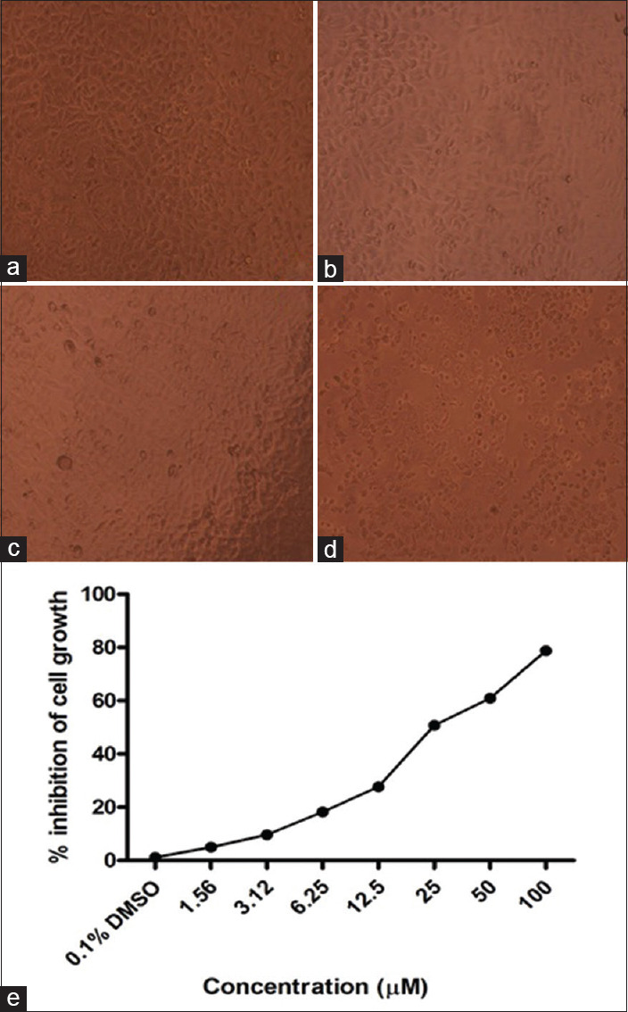
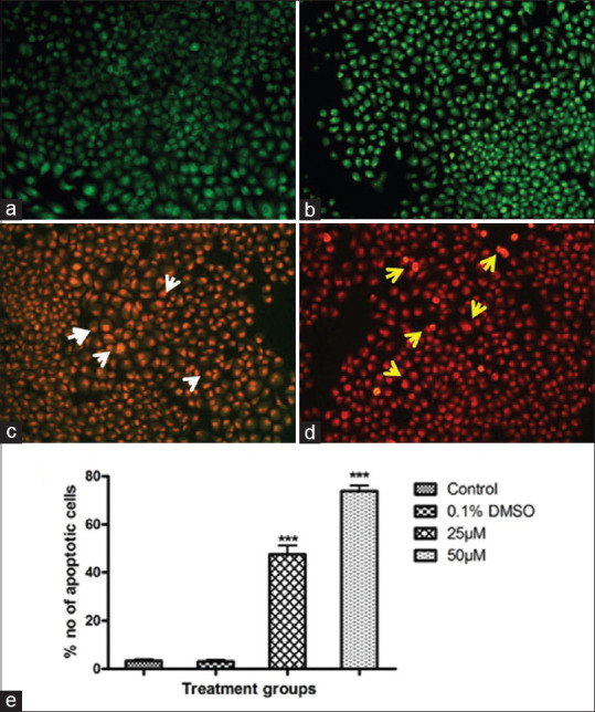
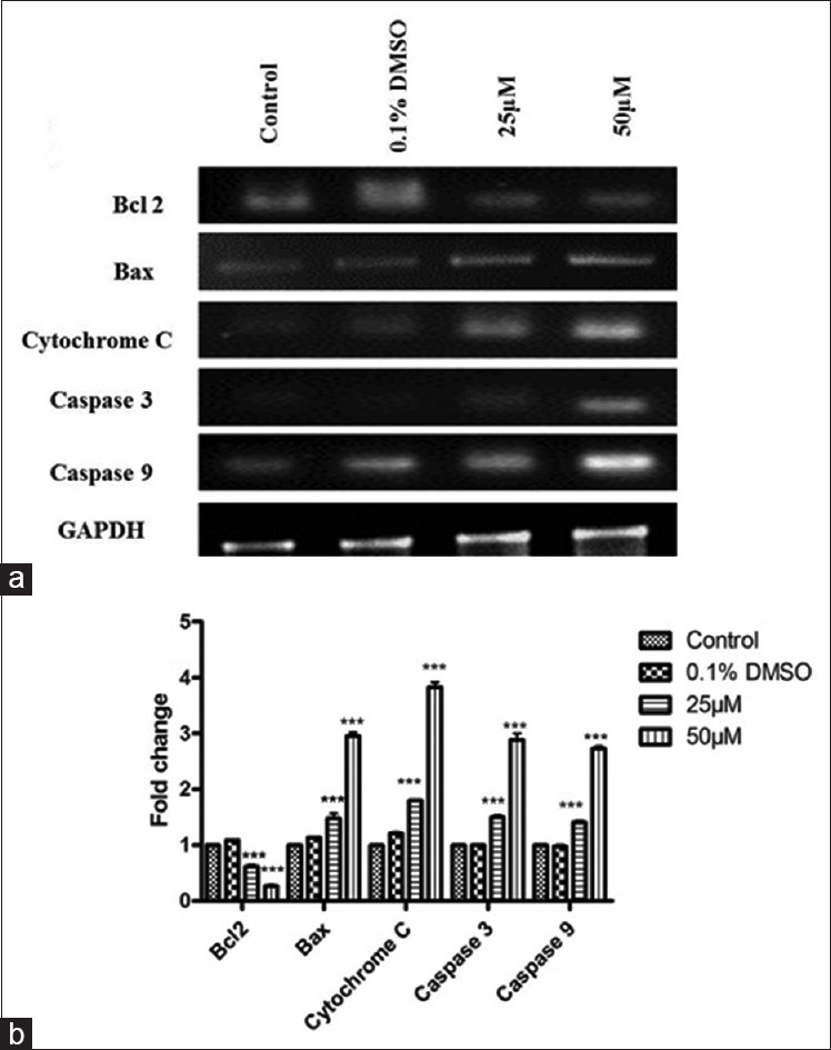
Cytotoxic effect of SA was determined by 3-(4,5-dimethylthiazol-2-yl)-2,5-diphenylte trazolium bromide assay, using concentrations of 25 and 50 μM/mL for 24 h. At the end of the treatment period, apoptotic markers such as caspase 3 and 9, bcl-2, bax and cytochrome c were evaluated by semiquantitative reverse transcription-polymerase chain reaction. SA-induced morphological changes were investigated by acridine orange/ethidium bromide dual staining.
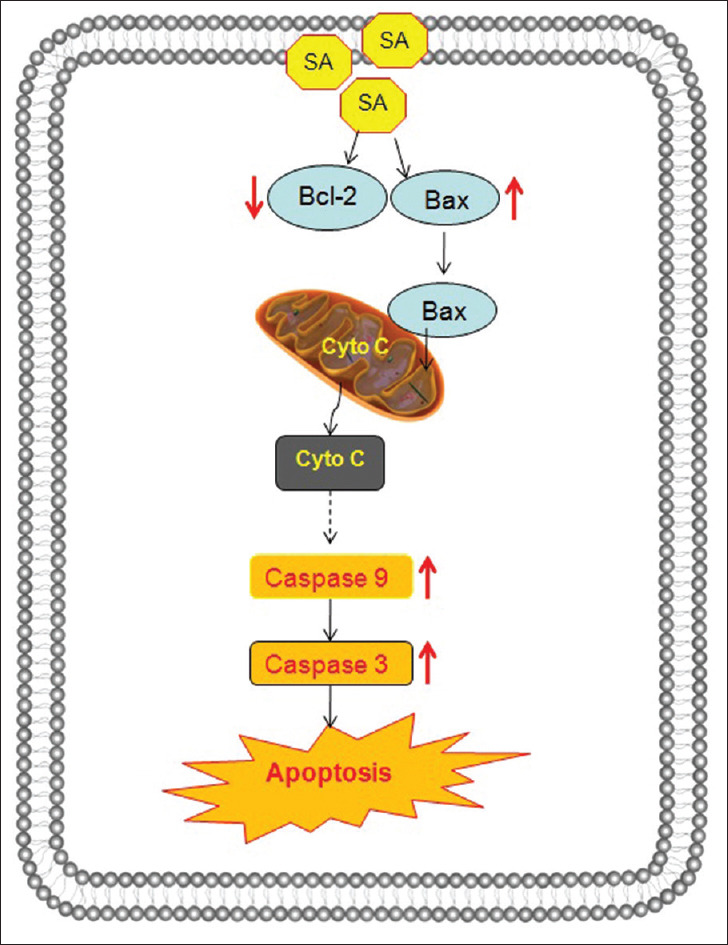
SA inhibited the proliferation and induced cytotoxicity in SCC-25 cells in a concentration-dependent manner. SA treatment caused apoptosis-related morphological changes as evidenced by the dual staining and the modulation of apoptotic marker gene expressions. SA treatments modulated bcl-2/bax homeostasis and increased the expressions of cytochrome c and caspases 3 and 9.
SA specifically induces cell death and inhibits the proliferation in OSCC cells through intrinsic/mitochondrial apoptosis pathway, suggesting that SA may be an effective agent for the treatment of human OSCC.
丁香酸(SA)长期以来一直被用作传统药物,已知具有抗氧化、保肝、神经保护和抗癌作用。关于SA对鳞状癌细胞(SCC)-25(一种人类口腔鳞状细胞癌(OSCC)细胞系)的抗癌作用尚未见研究报道。
本研究旨在评估SA对SCC-25细胞的细胞毒性潜力。
采用3-(4,5-二甲基噻唑-2-基)-2,5-二苯基四氮唑溴盐法测定SA的细胞毒性,使用浓度为25和50 μM/mL处理24小时。在处理期结束时,通过半定量逆转录-聚合酶链反应评估凋亡标志物如半胱天冬酶3和9、bcl-2、bax和细胞色素c。通过吖啶橙/溴化乙锭双重染色研究SA诱导的形态学变化。
SA以浓度依赖性方式抑制SCC-25细胞的增殖并诱导细胞毒性。双重染色和凋亡标志物基因表达的调节证明SA处理导致了与凋亡相关的形态学变化。SA处理调节了bcl-2/bax平衡,并增加了细胞色素c以及半胱天冬酶3和9的表达。
SA通过内源性/线粒体凋亡途径特异性诱导OSCC细胞死亡并抑制其增殖,提示SA可能是治疗人类OSCC的有效药物。