Lemonnier Tom, Dupré Aude, Jessus Catherine
Laboratoire de Biologie du Développement-Institut de Biologie Paris Seine, LBD-IBPS, Sorbonne Université, CNRS, 75005 Paris, France.
Cell Div. 2020 May 25;15:9. doi: 10.1186/s13008-020-00065-2. eCollection 2020.
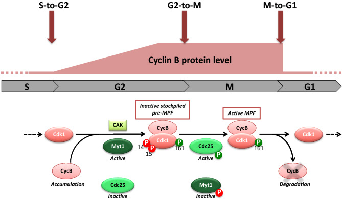
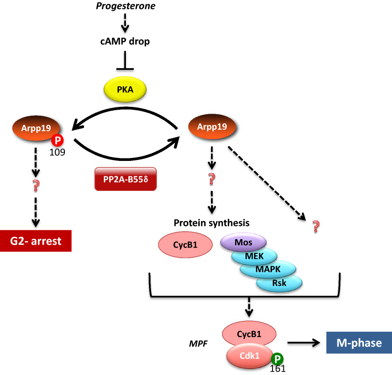
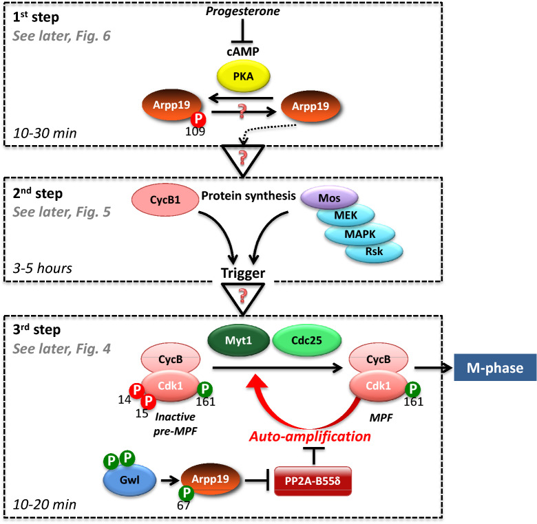
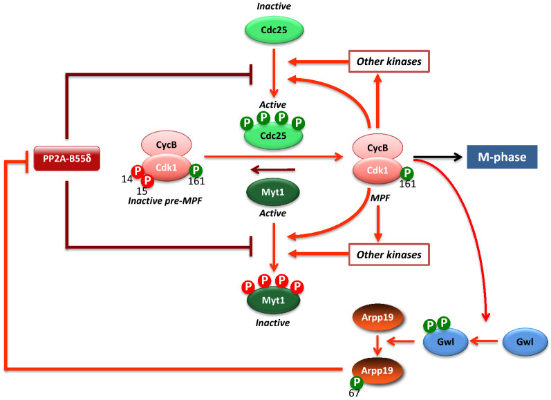
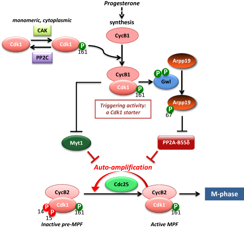
Cell division is orchestrated by the phosphorylation and dephosphorylation of thousands of proteins. These post-translational modifications underlie the molecular cascades converging to the activation of the universal mitotic kinase, Cdk1, and entry into cell division. They also govern the structural events that sustain the mechanics of cell division. While the role of protein kinases in mitosis has been well documented by decades of investigations, little was known regarding the control of protein phosphatases until the recent years. However, the regulation of phosphatase activities is as essential as kinases in controlling the activation of Cdk1 to enter M-phase. The regulation and the function of phosphatases result from post-translational modifications but also from the combinatorial association between conserved catalytic subunits and regulatory subunits that drive their substrate specificity, their cellular localization and their activity. It now appears that sequential dephosphorylations orchestrated by a network of phosphatase activities trigger Cdk1 activation and then order the structural events necessary for the timely execution of cell division. This review discusses a series of recent works describing the important roles played by protein phosphatases for the proper regulation of meiotic division. Many breakthroughs in the field of cell cycle research came from studies on oocyte meiotic divisions. Indeed, the meiotic division shares most of the molecular regulators with mitosis. The natural arrests of oocytes in G2 and in M-phase, the giant size of these cells, the variety of model species allowing either biochemical or imaging as well as genetics approaches explain why the process of meiosis has served as an historical model to decipher signalling pathways involved in the G2-to-M transition. The review especially highlights how the phosphatase PP2A-B55δ critically orchestrates the timing of meiosis resumption in amphibian oocytes. By opposing the kinase PKA, PP2A-B55δ controls the release of the G2 arrest through the dephosphorylation of their substrate, Arpp19. Few hours later, the inhibition of PP2A-B55δ by Arpp19 releases its opposing kinase, Cdk1, and triggers M-phase. In coordination with a variety of phosphatases and kinases, the PP2A-B55δ/Arpp19 duo therefore emerges as the key effector of the G2-to-M transition.
细胞分裂由数千种蛋白质的磷酸化和去磷酸化过程精心调控。这些翻译后修饰构成了汇聚至通用有丝分裂激酶Cdk1激活及进入细胞分裂过程的分子级联反应的基础。它们还掌控着维持细胞分裂机制的结构事件。尽管数十年来的研究已充分证明蛋白激酶在有丝分裂中的作用,但直到近年来,人们对蛋白磷酸酶的调控了解甚少。然而,磷酸酶活性的调节在控制Cdk1激活以进入M期方面与激酶同样重要。磷酸酶的调节和功能不仅源于翻译后修饰,还源于保守催化亚基与调节亚基之间的组合关联,这些关联决定了它们的底物特异性、细胞定位及其活性。现在看来,由磷酸酶活性网络精心编排的顺序去磷酸化触发了Cdk1的激活,进而安排了及时执行细胞分裂所需的结构事件。本综述讨论了一系列近期研究,这些研究描述了蛋白磷酸酶在减数分裂正确调控中所起的重要作用。细胞周期研究领域的许多突破都来自对卵母细胞减数分裂的研究。事实上,减数分裂与有丝分裂共享大部分分子调节因子。卵母细胞在G2期和M期的自然停滞、这些细胞的巨大尺寸、允许进行生化、成像以及遗传学研究的多种模式物种,解释了为什么减数分裂过程一直是破译参与G2到M期转换的信号通路的历史模型。该综述特别强调了磷酸酶PP2A - B55δ如何在两栖类卵母细胞中严格协调减数分裂恢复的时间。通过对抗激酶PKA,PP2A - B55δ通过使其底物Arpp19去磷酸化来控制G2期停滞的解除。几小时后,Arpp19对PP2A - B55δ的抑制作用释放了其对抗激酶Cdk1,并触发M期。因此,与多种磷酸酶和激酶协同作用,PP2A - B55δ/Arpp19二元组合成为G2到M期转换的关键效应器。