Morii Chiharu, Tanaka Hiroyoshi Y, Izushi Yasuhisa, Nakao Natsumi, Yamamoto Masaya, Matsubara Hiromi, Kano Mitsunobu R, Ogawa Aiko
Department of Pharmaceutical Biomedicine, Graduate School of Medicine, Dentistry, and Pharmaceutical Sciences, Okayama University, Okayama, Japan.
Division of Molecular and Cellular Medicine, Department of Clinical Science, National Hospital Organization Okayama Medical Center, Okayama, Japan.
Front Bioeng Biotechnol. 2020 May 20;8:482. doi: 10.3389/fbioe.2020.00482. eCollection 2020.
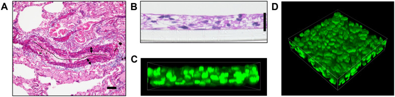
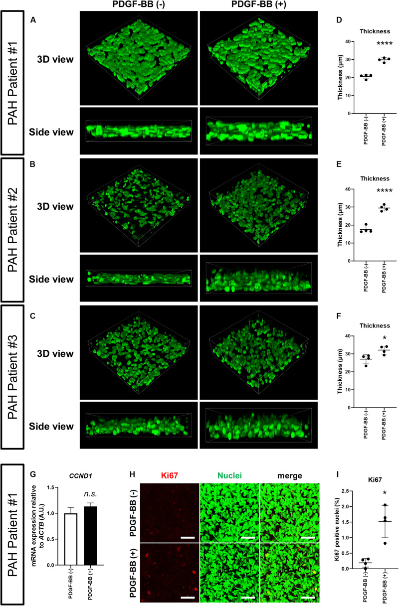
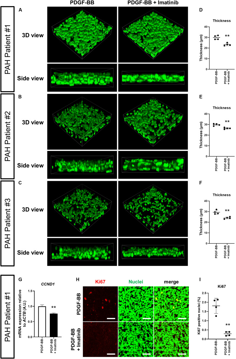
In pulmonary arterial hypertension (PAH), excessive proliferation of pulmonary artery smooth muscle cells (PASMCs) causes vascular medial thickening. Medial thickening is a histopathological hallmark of pulmonary vascular remodeling, the central disease process driving PAH progression. Pulmonary vascular remodeling causes stenosis and/or obstruction of small pulmonary arteries. This leads to increased pulmonary vascular resistance, elevated pulmonary arterial pressure, and ultimately right heart failure. To improve the survival of PAH patients, which remains at approximately 60% at 3 years after diagnosis, the development of novel PAH-targeted drugs is desired. To this end, a detailed understanding of the mechanisms underlying excessive PASMC proliferation and the medial thickening that ensues is necessary. However, a lack of models that recapitulate medial thickening impedes our deeper understanding of the pathogenetic mechanisms involved. In the present study, we applied 3-dimensional (3D) cell culture technology to develop a novel model of the pulmonary artery medial layer using human PAH patient-derived PASMCs. The addition of platelet-derived growth factor (PDGF)-BB, a mitogen known to promote excessive PASMC proliferation in PAH, resulted in increased thickness of the 3D-PAH media tissues. Conversely, administration of the PDGF receptor inhibitor imatinib or other clinical PAH drugs inhibited this medial thickening-inducing effect of PDGF-BB. Altogether, by using 3D cell culture technology, we report the generation of an model of medial thickening in PAH, which had hitherto not been successfully modeled . This model is potentially useful for assessing the ability of candidate PAH drugs to suppress medial thickening.
在肺动脉高压(PAH)中,肺动脉平滑肌细胞(PASMCs)过度增殖导致血管中层增厚。中层增厚是肺血管重塑的组织病理学标志,是推动PAH进展的核心疾病过程。肺血管重塑导致小肺动脉狭窄和/或阻塞。这会导致肺血管阻力增加、肺动脉压力升高,最终导致右心衰竭。为了提高PAH患者的生存率(诊断后3年生存率仍约为60%),需要开发新型PAH靶向药物。为此,有必要详细了解PASMC过度增殖及随之而来的中层增厚的潜在机制。然而,缺乏能够重现中层增厚的模型阻碍了我们对相关发病机制的深入理解。在本研究中,我们应用三维(3D)细胞培养技术,利用源自PAH患者的PASMCs开发了一种新型的肺动脉中层模型。添加血小板衍生生长因子(PDGF)-BB(一种已知可促进PAH中PASMC过度增殖的促有丝分裂原)导致3D-PAH中层组织厚度增加。相反,给予PDGF受体抑制剂伊马替尼或其他临床PAH药物可抑制PDGF-BB的这种中层增厚诱导作用。总之,通过使用3D细胞培养技术,我们报道了一种PAH中层增厚模型的建立,这是迄今为止尚未成功建立的模型。该模型可能有助于评估候选PAH药物抑制中层增厚的能力。