• 文献检索
  • 文档翻译
  • 深度研究
  • 学术资讯
  • Suppr Zotero 插件Zotero 插件
  • 邀请有礼
  • 套餐&价格
  • 历史记录
应用&插件
Suppr Zotero 插件Zotero 插件浏览器插件Mac 客户端Windows 客户端微信小程序
定价
高级版会员购买积分包购买API积分包
服务
文献检索文档翻译深度研究API 文档MCP 服务
关于我们
关于 Suppr公司介绍联系我们用户协议隐私条款
关注我们

Suppr 超能文献

核心技术专利:CN118964589B侵权必究
粤ICP备2023148730 号-1Suppr @ 2026

文献检索

告别复杂PubMed语法,用中文像聊天一样搜索,搜遍4000万医学文献。AI智能推荐,让科研检索更轻松。

立即免费搜索

文件翻译

保留排版,准确专业,支持PDF/Word/PPT等文件格式,支持 12+语言互译。

免费翻译文档

深度研究

AI帮你快速写综述,25分钟生成高质量综述,智能提取关键信息,辅助科研写作。

立即免费体验

磁共振成像和扩散峰度成像揭示的豚鼠出生后脑发育

Postnatal Guinea Pig Brain Development, as Revealed by Magnetic Resonance and Diffusion Kurtosis Imaging.

作者信息

Mullins Roger J, Xu Su, Zhuo Jiachen, Roys Steve, Pereira Edna F R, Albuquerque Edson X, Gullapalli Rao P

机构信息

Department of Diagnostic Radiology & Nuclear Medicine, University of Maryland School of Medicine, Baltimore, MD 21201, USA.

Program in Neuroscience, University of Maryland School of Medicine, Baltimore, MD 21201, USA.

出版信息

Brain Sci. 2020 Jun 12;10(6):365. doi: 10.3390/brainsci10060365.

DOI:10.3390/brainsci10060365
PMID:32545593
原文链接:https://pmc.ncbi.nlm.nih.gov/articles/PMC7349860/
Abstract

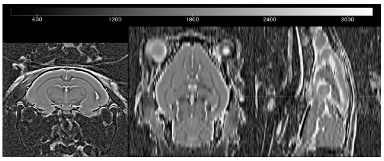
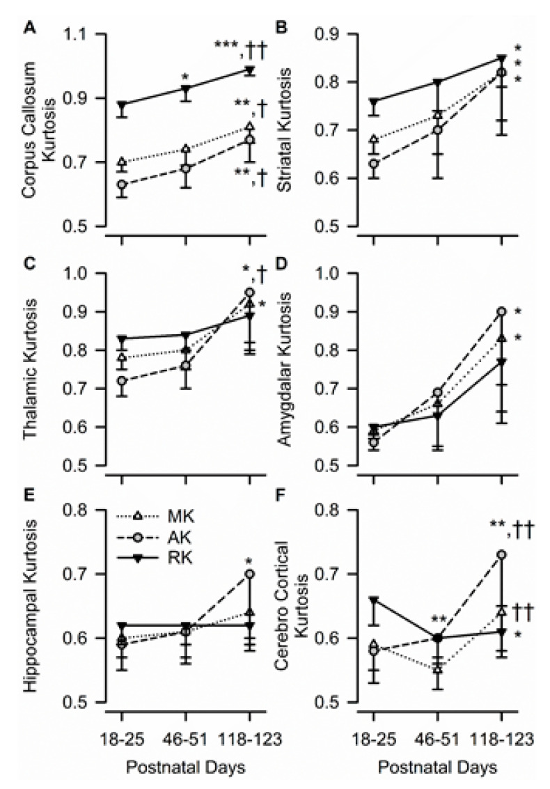
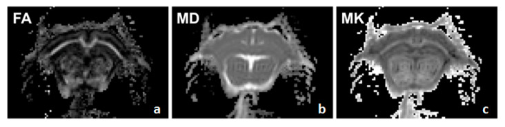
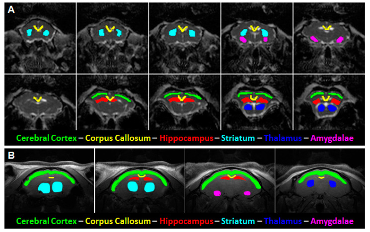
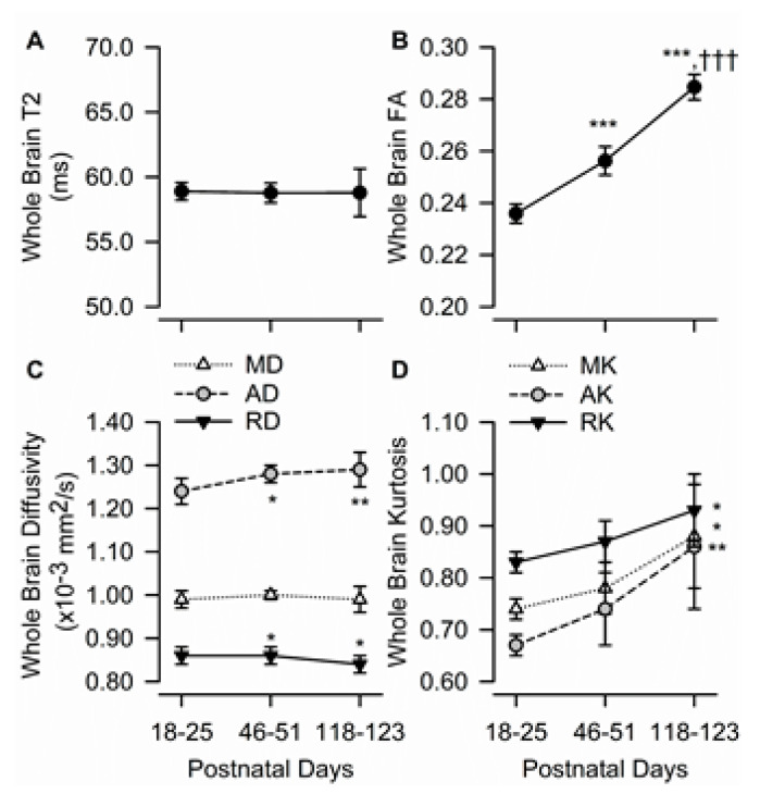
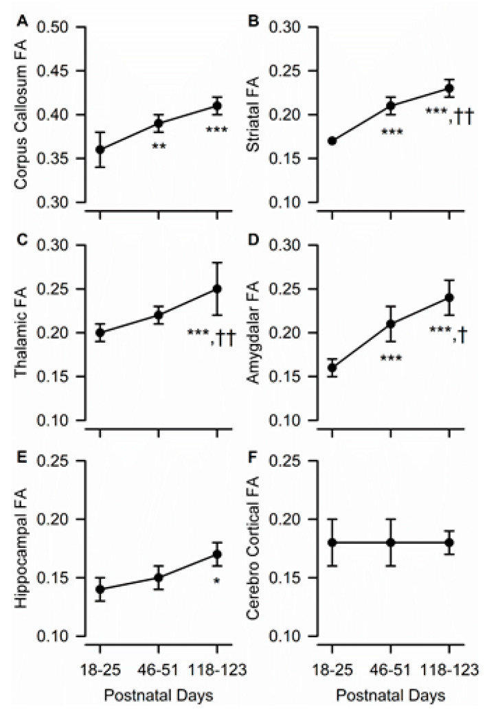
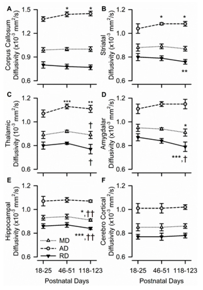
This study used magnetic resonance imaging (MRI) to identify age dependent brain structural characteristics in Dunkin Hartley guinea pigs. Anatomical T-weighted images, diffusion kurtosis (DKI) imaging, and T relaxometry measures were acquired from a cohort of male guinea pigs from postnatal day (PND) 18-25 (juvenile) to PND 46-51 (adolescent) and PND 118-123 (young adult). Whole-brain diffusion measures revealed the distinct effects of maturation on the microstructural complexity of the male guinea pig brain. Specifically, fractional anisotropy (FA), as well as mean, axial, and radial kurtosis in the corpus callosum, amygdala, dorsal-ventral striatum, and thalamus significantly increased from PND 18-25 to PND 118-123. Age-related alterations in DKI measures within these brain regions paralleled the overall alterations observed in the whole brain. Age-related changes in FA and kurtosis in the gray matter-dominant parietal cerebral cortex and dorsal hippocampus were less pronounced than in the other brain regions. The regional data analysis revealed that between-age changes of diffusion kurtosis metrics were more pronounced than those observed in diffusion tensor metrics. The age-related anatomical differences reported here may be important determinants of the age-dependent neurobehavior of guinea pigs in different tasks.

摘要

本研究使用磁共振成像(MRI)来确定邓金·哈特利豚鼠大脑结构特征的年龄依赖性。从一组雄性豚鼠中获取了解剖T加权图像、扩散峰度成像(DKI)和T弛豫测量值,这些豚鼠的年龄从出生后第18 - 25天(幼年)到第46 - 51天(青春期)以及第118 - 123天(青年期)。全脑扩散测量揭示了成熟对雄性豚鼠大脑微观结构复杂性的不同影响。具体而言,胼胝体、杏仁核、背腹侧纹状体和丘脑的分数各向异性(FA)以及平均、轴向和径向峰度从出生后第18 - 25天到第118 - 123天显著增加。这些脑区内DKI测量值的年龄相关变化与全脑观察到的总体变化相似。灰质占主导的顶叶大脑皮层和背侧海马体中FA和峰度的年龄相关变化不如其他脑区明显。区域数据分析表明,扩散峰度指标的年龄间变化比扩散张量指标更明显。这里报告的年龄相关解剖差异可能是豚鼠在不同任务中年龄依赖性神经行为的重要决定因素。

https://cdn.ncbi.nlm.nih.gov/pmc/blobs/fd4b/7349860/a3c47df565a1/brainsci-10-00365-g005.jpg
https://cdn.ncbi.nlm.nih.gov/pmc/blobs/fd4b/7349860/8b06df7751dd/brainsci-10-00365-g0A1.jpg
https://cdn.ncbi.nlm.nih.gov/pmc/blobs/fd4b/7349860/d24a13c20750/brainsci-10-00365-g0A2.jpg
https://cdn.ncbi.nlm.nih.gov/pmc/blobs/fd4b/7349860/4380bbf3e5fe/brainsci-10-00365-g001.jpg
https://cdn.ncbi.nlm.nih.gov/pmc/blobs/fd4b/7349860/a9a01679d5bb/brainsci-10-00365-g002.jpg
https://cdn.ncbi.nlm.nih.gov/pmc/blobs/fd4b/7349860/e19b4c0e98fc/brainsci-10-00365-g003.jpg
https://cdn.ncbi.nlm.nih.gov/pmc/blobs/fd4b/7349860/92684aa974ce/brainsci-10-00365-g004.jpg
https://cdn.ncbi.nlm.nih.gov/pmc/blobs/fd4b/7349860/a3c47df565a1/brainsci-10-00365-g005.jpg

相似文献

1
Postnatal Guinea Pig Brain Development, as Revealed by Magnetic Resonance and Diffusion Kurtosis Imaging.磁共振成像和扩散峰度成像揭示的豚鼠出生后脑发育
Brain Sci. 2020 Jun 12;10(6):365. doi: 10.3390/brainsci10060365.
2
Application of diffusion kurtosis imaging in neonatal brain development.扩散峰度成像在新生儿脑发育中的应用。
Front Pediatr. 2023 Mar 27;11:1112121. doi: 10.3389/fped.2023.1112121. eCollection 2023.
3
Differences in Gaussian diffusion tensor imaging and non-Gaussian diffusion kurtosis imaging model-based estimates of diffusion tensor invariants in the human brain.基于高斯扩散张量成像和非高斯扩散峰度成像模型的人脑扩散张量不变量估计差异。
Med Phys. 2016 May;43(5):2464. doi: 10.1118/1.4946819.
4
Can diffusion kurtosis imaging improve the sensitivity and specificity of detecting microstructural alterations in brain tissue chronically after experimental stroke? Comparisons with diffusion tensor imaging and histology.弥散峰度成像能否提高实验性中风后慢性脑组织微观结构改变的检测敏感性和特异性?与弥散张量成像和组织学的比较。
Neuroimage. 2014 Aug 15;97:363-73. doi: 10.1016/j.neuroimage.2014.04.013. Epub 2014 Apr 15.
5
Microstructural brain abnormalities correlate with neurocognitive dysfunction in minimal hepatic encephalopathy: a diffusion kurtosis imaging study.脑微结构异常与轻微肝性脑病的神经认知功能障碍相关:一项扩散峰度成像研究
Neuroradiology. 2019 Jun;61(6):685-694. doi: 10.1007/s00234-019-02201-4. Epub 2019 Mar 27.
6
Diffusion kurtosis imaging allows the early detection and longitudinal follow-up of amyloid-β-induced pathology.弥散峰度成像可实现对淀粉样-β诱导的病变的早期检测和纵向随访。
Alzheimers Res Ther. 2018 Jan 9;10(1):1. doi: 10.1186/s13195-017-0329-8.
7
Microstructural alterations of cortical and deep gray matter over a season of high school football revealed by diffusion kurtosis imaging.弥散峰度成像显示,高中橄榄球赛季中皮质和深部灰质的微观结构改变。
Neurobiol Dis. 2018 Nov;119:79-87. doi: 10.1016/j.nbd.2018.07.020. Epub 2018 Jul 23.
8
Performances of diffusion kurtosis imaging and diffusion tensor imaging in detecting white matter abnormality in schizophrenia.扩散峰度成像和扩散张量成像在检测精神分裂症白质异常中的表现。
Neuroimage Clin. 2014 Dec 9;7:170-6. doi: 10.1016/j.nicl.2014.12.008. eCollection 2015.
9
[Diffusion kurtosis imaging in diffuse axonal injury].[扩散峰度成像在弥漫性轴索损伤中的应用]
Zh Vopr Neirokhir Im N N Burdenko. 2019;83(3):5-16. doi: 10.17116/neiro2019830315.
10
Evaluation of patients with relapsing-remitting multiple sclerosis using tract-based spatial statistics analysis: diffusion kurtosis imaging.使用基于纤维束的空间统计学分析评估复发缓解型多发性硬化症患者:扩散峰度成像
BMC Neurol. 2018 Aug 7;18(1):108. doi: 10.1186/s12883-018-1108-2.

引用本文的文献

1
Dual isolation of primary neurons and oligodendrocytes from guinea pig frontal cortex.从豚鼠额叶皮质双重分离原代神经元和少突胶质细胞。
Front Cell Neurosci. 2024 Jan 10;17:1298685. doi: 10.3389/fncel.2023.1298685. eCollection 2023.
2
Brain structure analysis of different age groups of Diannan small-ear pigs.滇南小耳猪不同年龄组的脑结构分析
Ibrain. 2022 Aug 13;8(3):314-323. doi: 10.1002/ibra.12058. eCollection 2022 Fall.
3
Olfactory dysfunction in aging and neurodegenerative diseases.衰老和神经退行性疾病中的嗅觉功能障碍。

本文引用的文献

1
Characterization of Alzheimer's Disease Using Ultra-high b-values Apparent Diffusion Coefficient and Diffusion Kurtosis Imaging.使用超高b值表观扩散系数和扩散峰度成像对阿尔茨海默病进行特征描述。
Aging Dis. 2019 Oct 1;10(5):1026-1036. doi: 10.14336/AD.2018.1129. eCollection 2019 Oct.
2
Diffusion Kurtosis Imaging Detects Microstructural Changes in a Methamphetamine-Induced Mouse Model of Parkinson's Disease.扩散峰度成像检测到甲基苯丙胺诱导的帕金森病小鼠模型中的微观结构变化。
Neurotox Res. 2019 Nov;36(4):724-735. doi: 10.1007/s12640-019-00068-0. Epub 2019 Jun 18.
3
Longitudinal Microstructural Changes in Traumatic Brain Injury in Rats: A Diffusional Kurtosis Imaging, Histology, and Behavior Study.
Ageing Res Rev. 2021 Sep;70:101416. doi: 10.1016/j.arr.2021.101416. Epub 2021 Jul 27.
大鼠创伤性脑损伤的纵向微观结构变化:弥散峰度成像、组织学和行为研究。
AJNR Am J Neuroradiol. 2018 Sep;39(9):1650-1656. doi: 10.3174/ajnr.A5737. Epub 2018 Jul 26.
4
Guinea pig models for translation of the developmental origins of health and disease hypothesis into the clinic.豚鼠模型将健康与疾病起源假说的发育翻译转化为临床实践。
J Physiol. 2018 Dec;596(23):5535-5569. doi: 10.1113/JP274948. Epub 2018 May 30.
5
Exposure of Developing Brain to General Anesthesia: What Is the Animal Evidence?发育中大脑接触全身麻醉:动物研究有何证据?
Anesthesiology. 2018 Apr;128(4):832-839. doi: 10.1097/ALN.0000000000002047.
6
Developmental neurotoxicity of the organophosphorus insecticide chlorpyrifos: from clinical findings to preclinical models and potential mechanisms.有机磷杀虫剂毒死蜱的发育神经毒性:从临床发现到临床前模型及潜在机制
J Neurochem. 2017 Aug;142 Suppl 2(Suppl 2):162-177. doi: 10.1111/jnc.14077.
7
Diffusion Kurtosis Imaging Detects Microstructural Changes in the Brain after Acute Alcohol Intoxication in Rats.扩散峰度成像检测大鼠急性酒精中毒后脑内微观结构变化。
Biomed Res Int. 2017;2017:4757025. doi: 10.1155/2017/4757025. Epub 2017 Jan 18.
8
Early and progressive microstructural brain changes in mice overexpressing human α-Synuclein detected by diffusion kurtosis imaging.通过扩散峰度成像检测过度表达人α-突触核蛋白的小鼠早期和进行性的微观结构脑变化。
Brain Behav Immun. 2017 Mar;61:197-208. doi: 10.1016/j.bbi.2016.11.027. Epub 2016 Dec 5.
9
Repetitive Model of Mild Traumatic Brain Injury Produces Cortical Abnormalities Detectable by Magnetic Resonance Diffusion Imaging, Histopathology, and Behavior.轻度创伤性脑损伤的重复模型会产生可通过磁共振扩散成像、组织病理学和行为检测到的皮质异常。
J Neurotrauma. 2017 Apr 1;34(7):1364-1381. doi: 10.1089/neu.2016.4569. Epub 2016 Dec 20.
10
Genetic variants associated with neurodegenerative Alzheimer disease in natural models.自然模型中与神经退行性阿尔茨海默病相关的基因变异。
Biol Res. 2016 Feb 26;49:14. doi: 10.1186/s40659-016-0072-9.