Minami Teruya, Terada Takuro, Mitsui Takeshi, Nakanuma Yasuni
Department of Surgery, Fukui-ken Saiseikai Hospital, Fukui, Japan.
Department of Pathology, Fukui-ken Saiseikai Hospital, Fukui, Japan.
Surg Case Rep. 2020 Jun 18;6(1):141. doi: 10.1186/s40792-020-00903-z.
Heterotopic pancreas (HP) is defined as pancreatic tissue in organs with no anatomical continuity with the orthotopic pancreas. Based on the number of cases reported in the literature between the year 2000 and 2020, HP is rarely found causing malignant transformation of the duodenum. We herein report a case of adenocarcinoma arising from the HP in the first portion of the duodenum.
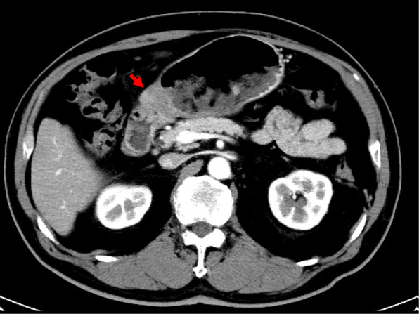
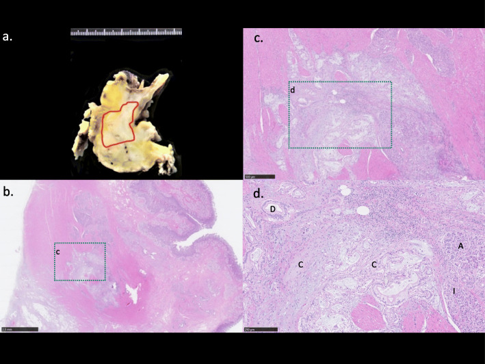
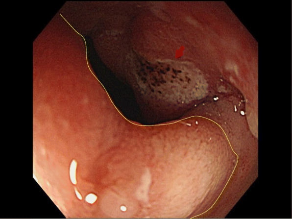
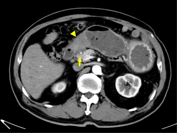
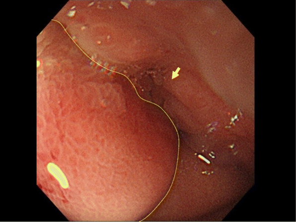
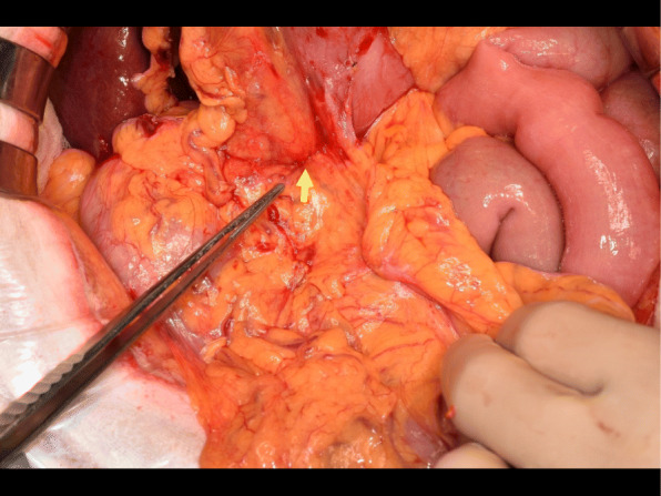
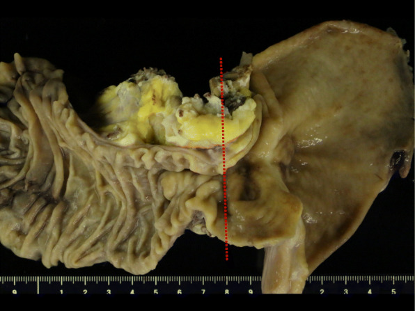
A 77-year-old Japanese man presented to our hospital with epigastric pain. Despite having undergone laparoscopic surgery for early sigmoid colon cancer a month earlier, serum levels of tumor-specific antigens, such as CA19-9, were elevated. After undergoing a series of radiologic examinations, the first portion of the duodenum was found thickened. However, a biopsy of the lesion showed no malignancy. Four months later, follow-up computed tomography (CT) scans showed that the lesion was thicker and involved the gastroduodenal artery (GDA), suggesting tumor invasion. A new biopsy did not detect the malignancy. However, serum tumor-specific antigen levels increased, especially duke pancreatic monoclonal antigen type 2 (5287 U/mL), in the absence of tumor in the orthotopic pancreas. The follow-up CT imaging showed a malignant tumor in the first portion of the duodenum. Five months later, we performed a subtotal stomach-preserving pancreaticoduodenectomy (SSPPD) for duodenal or HP cancer in the first portion of the duodenum, finding a lesion from the pyloric bulbs to the first portion of the duodenum, which invaded the adjacent pancreas and GDA. The pathological examination of the specimens revealed adenocarcinoma arising from HP. Nine months after surgery, no recurrence was found by radiologic imaging or tumor-specific antigen laboratory testing.
HP adenocarcinoma is rare and difficult to diagnose preoperatively due to its submucosal location. Therefore, a careful follow-up with blood testing and radiologic imaging, as well as diagnostic surgery, is recommended.
异位胰腺(HP)是指位于与正常胰腺无解剖学连续性的器官内的胰腺组织。根据2000年至2020年文献报道的病例数量,很少发现HP会导致十二指肠发生恶性转化。我们在此报告一例起源于十二指肠第一部HP的腺癌病例。
一名77岁的日本男性因上腹部疼痛就诊于我院。尽管一个月前因早期乙状结肠癌接受了腹腔镜手术,但血清肿瘤特异性抗原如CA19-9水平升高。经过一系列影像学检查,发现十二指肠第一部增厚。然而,病变活检未发现恶性肿瘤。四个月后,随访计算机断层扫描(CT)显示病变增厚并累及胃十二指肠动脉(GDA),提示肿瘤侵犯。再次活检未检测到恶性肿瘤。然而,在正常胰腺无肿瘤的情况下,血清肿瘤特异性抗原水平升高,尤其是杜克胰腺单克隆抗原2型(5287 U/mL)。随访CT成像显示十二指肠第一部有恶性肿瘤。五个月后,我们对十二指肠第一部的十二指肠或HP癌进行了保留胃的胰十二指肠次全切除术(SSPPD),发现从幽门球部到十二指肠第一部有一个病变,侵犯了相邻的胰腺和GDA。标本的病理检查显示为起源于HP的腺癌。术后九个月,影像学检查或肿瘤特异性抗原实验室检测均未发现复发。
HP腺癌罕见,因其位于黏膜下,术前难以诊断。因此,建议进行仔细的血液检测、影像学随访以及诊断性手术。