• 文献检索
  • 文档翻译
  • 深度研究
  • 学术资讯
  • Suppr Zotero 插件Zotero 插件
  • 邀请有礼
  • 套餐&价格
  • 历史记录
应用&插件
Suppr Zotero 插件Zotero 插件浏览器插件Mac 客户端Windows 客户端微信小程序
定价
高级版会员购买积分包购买API积分包
服务
文献检索文档翻译深度研究API 文档MCP 服务
关于我们
关于 Suppr公司介绍联系我们用户协议隐私条款
关注我们

Suppr 超能文献

核心技术专利:CN118964589B侵权必究
粤ICP备2023148730 号-1Suppr @ 2026

文献检索

告别复杂PubMed语法,用中文像聊天一样搜索,搜遍4000万医学文献。AI智能推荐,让科研检索更轻松。

立即免费搜索

文件翻译

保留排版,准确专业,支持PDF/Word/PPT等文件格式,支持 12+语言互译。

免费翻译文档

深度研究

AI帮你快速写综述,25分钟生成高质量综述,智能提取关键信息,辅助科研写作。

立即免费体验

内陆刺蜥( Pogona vitticeps )消化道的大体解剖学、组织学和血管分布。

Gross anatomy, histology and blood vessel topography of the alimentary canal of the Inland Bearded Dragon (Pogona vitticeps).

机构信息

Institute for Anatomy, University of Veterinary Medicine Hannover, Hannover, Germany.

Clinic for Small Mammals, Reptiles and Birds, University of Veterinary Medicine Hannover, Hannover, Germany.

出版信息

PLoS One. 2020 Jun 23;15(6):e0234736. doi: 10.1371/journal.pone.0234736. eCollection 2020.

DOI:10.1371/journal.pone.0234736
PMID:32574166
原文链接:https://pmc.ncbi.nlm.nih.gov/articles/PMC7310682/
Abstract

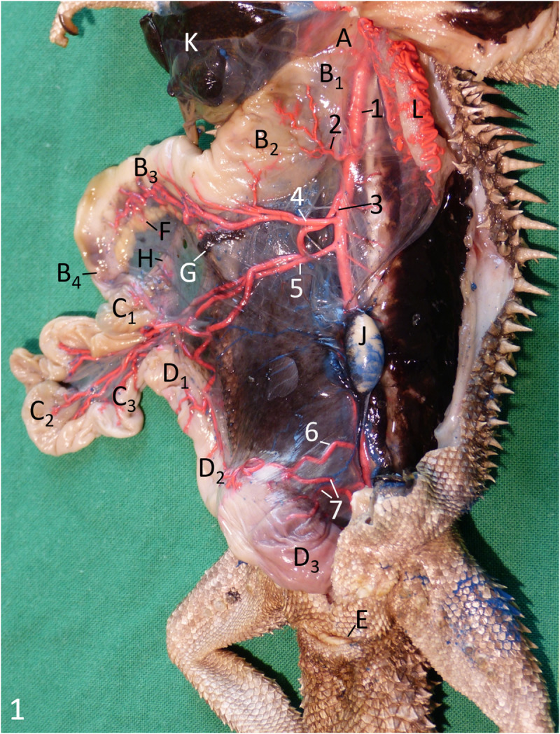
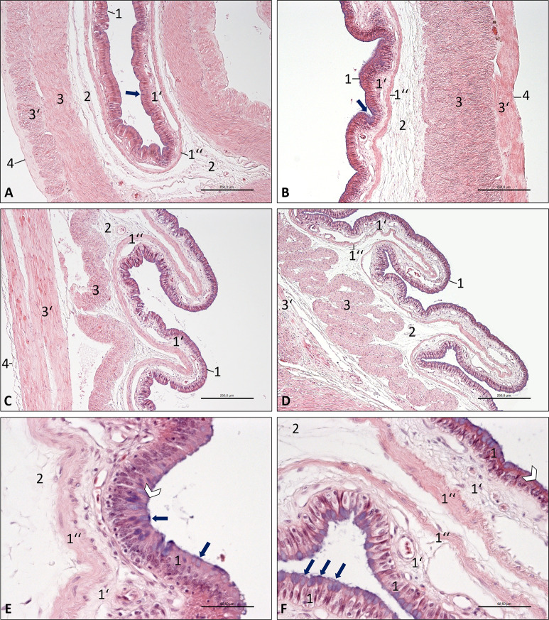
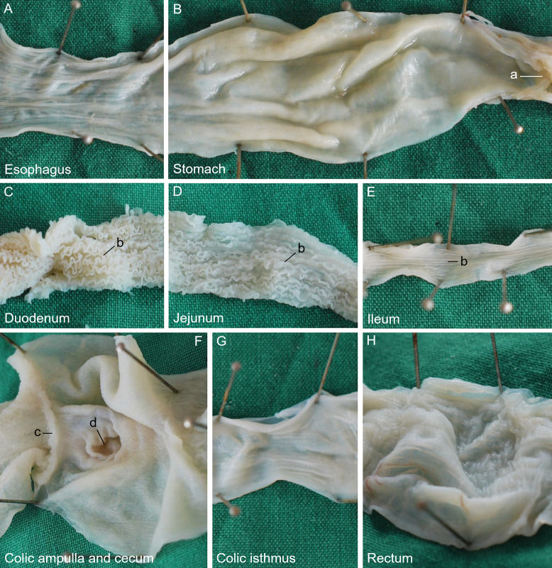
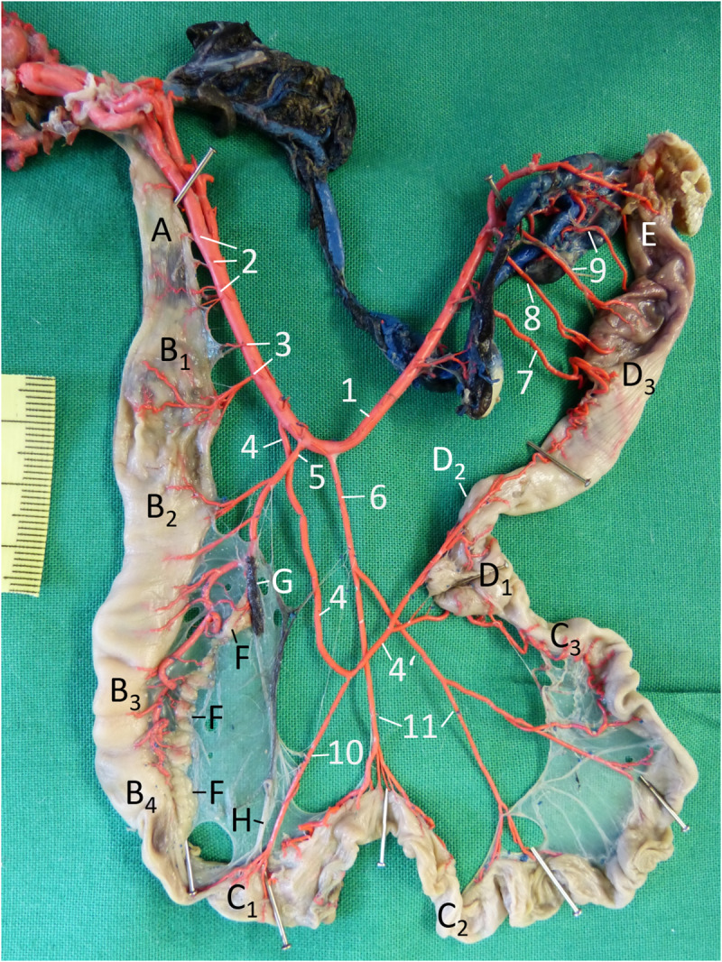
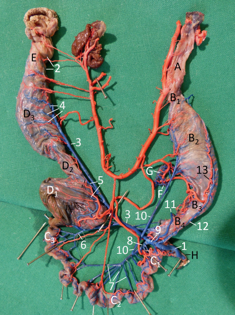
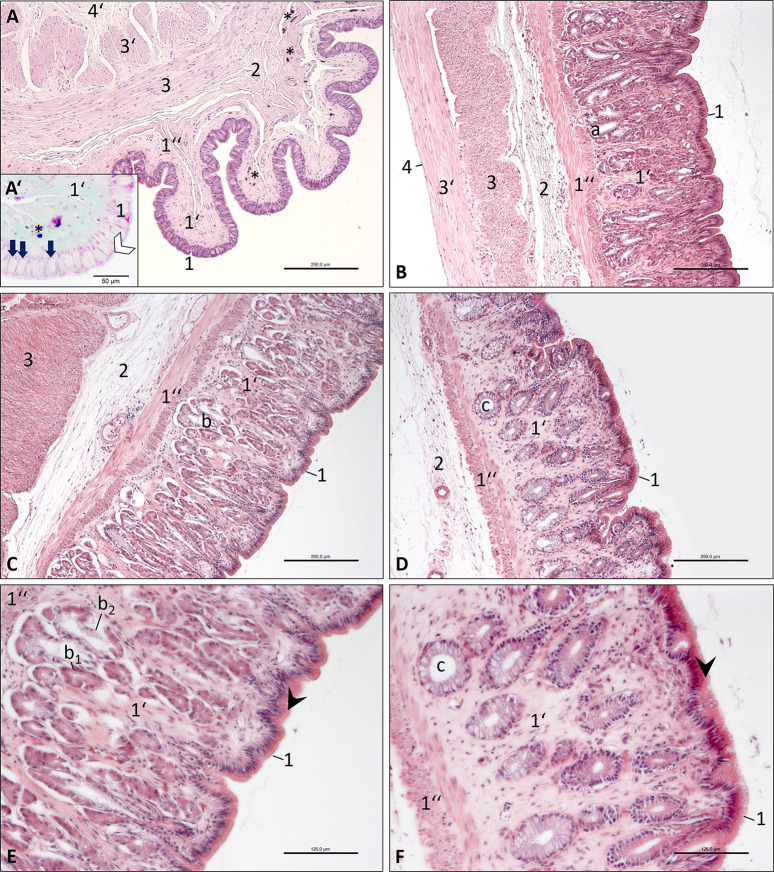
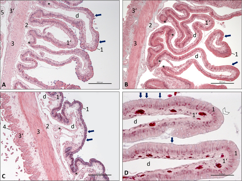
Imaging techniques have proved to be crucial for diagnosis in reptile species. The topography of the internal organs of bearded dragons has been described in recent studies as meeting the small animal practitioners´ demand for knowledge concerning their anatomy. However, the nomenclature in the respective literature is not uniform, which could lead to misunderstandings concerning the respective and/or affected parts of the alimentary canal. Therefore, the aim of this study was to provide clear information on anatomy and histology of the alimentary canal of bearded dragons including supplying blood vessels. For the dissection of the alimentary canal, 11 Inland Bearded Dragons (Pogona vitticeps) were used (five males, six females), which had been euthanised for clinical reasons other than those concerning the digestive tract or had died spontaneously. The supplying arteries were demonstrated by injecting red latex into the aorta, while the intestinal veins were filled with blue latex via the portal vein. Microscopic examination was carried out on specimens of seven additional bearded dragons using routine histologic procedures. Macroscopically, the sections of the alimentary canal from oral to aboral were distinguished into oesophagus, stomach, small intestine, colic ampulla, colic isthmus, rectum and cloaca. Differentiation of the duodenum, jejunum and ileum was only possible when considering the bile duct, the vasculature and the histology of the organ wall. Arteries supplying the oesophagus and the final straight part of the large intestine originated from the aorta in a segmental manner. Between these, three unpaired arteries arose from the aorta. Their branches supplied stomach and intestine excluding its last part. Based on the findings of the present study, a nomenclature for the different parts of the alimentary canal and the supplying blood vessels of bearded dragons is suggested which is well understandable for veterinary practitioners and is based on zoological knowledge of reptiles.

摘要

成像技术已被证明对爬行动物物种的诊断至关重要。最近的研究描述了鬃狮蜥内部器官的地形,满足了小动物从业者对其解剖结构知识的需求。然而,各自文献中的命名法并不统一,这可能导致对消化道的各个和/或受影响部分的误解。因此,本研究的目的是提供有关鬃狮蜥消化道的解剖学和组织学的明确信息,包括供应血管。为了解剖消化道,使用了 11 只内陆鬃狮蜥(Pogona vitticeps)(5 只雄性,6 只雌性),这些蜥蜥是由于与消化道无关的临床原因而被安乐死的,或者是自然死亡的。通过将红色乳胶注入主动脉来显示供应动脉,而通过门静脉将蓝色乳胶填充到肠静脉中。通过常规组织学程序对另外 7 只鬃狮蜥的标本进行了微观检查。宏观上,从口腔到肛门的消化道切片分为食道、胃、小肠、结肠壶腹、结肠峡部、直肠和泄殖腔。只有考虑到胆管、血管和器官壁的组织学,才能区分十二指肠、空肠和回肠。供应食道和大肠最后直段的动脉从主动脉呈节段性起源。在这些动脉之间,从主动脉发出了三条不成对的动脉。它们的分支供应胃和肠,不包括其最后部分。基于本研究的发现,建议为鬃狮蜥的不同消化道部分和供应血管提出一个命名法,该命名法对于兽医从业者来说是容易理解的,并且基于爬行动物的动物学知识。

https://cdn.ncbi.nlm.nih.gov/pmc/blobs/7ac4/7310682/b24928a65c60/pone.0234736.g007.jpg
https://cdn.ncbi.nlm.nih.gov/pmc/blobs/7ac4/7310682/1a8ef02a62c7/pone.0234736.g001.jpg
https://cdn.ncbi.nlm.nih.gov/pmc/blobs/7ac4/7310682/579be32aa0fe/pone.0234736.g002.jpg
https://cdn.ncbi.nlm.nih.gov/pmc/blobs/7ac4/7310682/68e59559b2d0/pone.0234736.g003.jpg
https://cdn.ncbi.nlm.nih.gov/pmc/blobs/7ac4/7310682/17aea0aa4d97/pone.0234736.g004.jpg
https://cdn.ncbi.nlm.nih.gov/pmc/blobs/7ac4/7310682/8b6b21db4bef/pone.0234736.g005.jpg
https://cdn.ncbi.nlm.nih.gov/pmc/blobs/7ac4/7310682/7a49a30d69e0/pone.0234736.g006.jpg
https://cdn.ncbi.nlm.nih.gov/pmc/blobs/7ac4/7310682/b24928a65c60/pone.0234736.g007.jpg

相似文献

1
Gross anatomy, histology and blood vessel topography of the alimentary canal of the Inland Bearded Dragon (Pogona vitticeps).内陆刺蜥( Pogona vitticeps )消化道的大体解剖学、组织学和血管分布。
PLoS One. 2020 Jun 23;15(6):e0234736. doi: 10.1371/journal.pone.0234736. eCollection 2020.
2
Specific anatomy and radiographic illustration of the digestive tract and transit time of two orally administered contrast media in Inland bearded dragons (Pogona vitticeps).内陆刺蜥( Pogona vitticeps )消化道的特定解剖结构和放射影像学表现,以及两种口服造影剂的通过时间。
PLoS One. 2019 Aug 22;14(8):e0221050. doi: 10.1371/journal.pone.0221050. eCollection 2019.
3
Ultrasonographic anatomy of bearded dragons (Pogona vitticeps).鬃狮蜥(鬃狮蜥)的超声解剖学
J Am Vet Med Assoc. 2015 Apr 15;246(8):868-76. doi: 10.2460/javma.246.8.868.
4
Ophthalmic examination, biometry, and histologic findings in captive inland bearded dragons (Pogona vitticeps).圈养内陆刺蜥( Pogona vitticeps )的眼科检查、生物测量和组织学发现。
Vet Ophthalmol. 2020 Jul;23(4):696-706. doi: 10.1111/vop.12779. Epub 2020 May 27.
5
Correction: Gross anatomy, histology and blood vessel topography of the alimentary canal of the Inland Bearded Dragon (Pogona vitticeps).更正:内陆鬃狮蜥(鬃狮蜥)消化道的大体解剖、组织学和血管形态学
PLoS One. 2024 Jul 18;19(7):e0307698. doi: 10.1371/journal.pone.0307698. eCollection 2024.
6
Radiographic anatomy and barium sulfate contrast transit time of the gastrointestinal tract of bearded dragons (Pogona vitticeps).鬃狮蜥(鬃狮蜥)胃肠道的放射解剖学及硫酸钡造影剂通过时间
Vet Radiol Ultrasound. 2014 May-Jun;55(3):241-50. doi: 10.1111/vru.12128.
7
Aneurysm Associated with Vascular Wall Degeneration in Bearded Dragons ().髯蜥血管壁退化相关的动脉瘤()。
8
Neurological examination in healthy adult inland bearded dragons (Pogona vitticeps).健康成年内陆刺蜥( Pogona vitticeps )的神经学检查。
J Am Vet Med Assoc. 2022 Apr 13;260(9):1013-1023. doi: 10.2460/javma.20.12.0706.
9
Pharmacokinetics of subcutaneously administered hydromorphone in bearded dragons (Pogona vitticeps) and red-eared slider turtles (Trachemys scripta elegans).皮下注射氢吗啡酮在鬃狮蜥(鬃狮蜥)和红耳龟(彩龟)体内的药代动力学。
Vet Anaesth Analg. 2019 May;46(3):352-359. doi: 10.1016/j.vaa.2018.12.002. Epub 2019 Jan 18.
10
First report of fatal disseminated microsporidiosis in two inland bearded dragons in Japan.日本两只内陆鬃狮蜥发生致命播散性微孢子虫病的首例报告。
JMM Case Rep. 2017 Apr 12;4(4):e005089. doi: 10.1099/jmmcr.0.005089. eCollection 2017 Apr.

引用本文的文献

1
Correction: Gross anatomy, histology and blood vessel topography of the alimentary canal of the Inland Bearded Dragon (Pogona vitticeps).更正:内陆鬃狮蜥(鬃狮蜥)消化道的大体解剖、组织学和血管形态学
PLoS One. 2024 Jul 18;19(7):e0307698. doi: 10.1371/journal.pone.0307698. eCollection 2024.

本文引用的文献

1
Specific anatomy and radiographic illustration of the digestive tract and transit time of two orally administered contrast media in Inland bearded dragons (Pogona vitticeps).内陆刺蜥( Pogona vitticeps )消化道的特定解剖结构和放射影像学表现,以及两种口服造影剂的通过时间。
PLoS One. 2019 Aug 22;14(8):e0221050. doi: 10.1371/journal.pone.0221050. eCollection 2019.
2
Ultrasonographic anatomy of bearded dragons (Pogona vitticeps).鬃狮蜥(鬃狮蜥)的超声解剖学
J Am Vet Med Assoc. 2015 Apr 15;246(8):868-76. doi: 10.2460/javma.246.8.868.
3
Radiographic anatomy and barium sulfate contrast transit time of the gastrointestinal tract of bearded dragons (Pogona vitticeps).
鬃狮蜥(鬃狮蜥)胃肠道的放射解剖学及硫酸钡造影剂通过时间
Vet Radiol Ultrasound. 2014 May-Jun;55(3):241-50. doi: 10.1111/vru.12128.
4
Comparative evaluation of the cadaveric and computed tomographic features of the coelomic cavity in the green iguana (Iguana iguana), black and white tegu (Tupinambis merianae) and bearded dragon (Pogona vitticeps).绿鬣蜥(Iguana iguana)、黑白泰加蜥(Tupinambis merianae)和鬃狮蜥(Pogona vitticeps)体腔的尸体解剖特征与计算机断层扫描特征的比较评估
Anat Histol Embryol. 2013 Dec;42(6):453-60. doi: 10.1111/ahe.12037. Epub 2013 Feb 14.
5
Gastrointestinal studies in the green iguana: technique and reference values.绿鬣蜥的胃肠道研究:技术与参考值
Vet Radiol Ultrasound. 2001 Nov-Dec;42(6):515-20. doi: 10.1111/j.1740-8261.2001.tb00979.x.