School of Computer Science, Tokyo University of Technology, 1401-1 Katakura-machi, Hachioji-shi, Tokyo 192-0982, Japan.
Graduate School of Sciences and Technology for Innovation, Yamaguchi University, 2-16-1 Tokiwadai, Ube-shi, Yamaguchi 755-8611, Japan.
Biomolecules. 2020 Jun 19;10(6):931. doi: 10.3390/biom10060931.
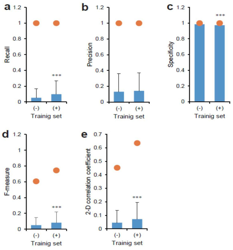
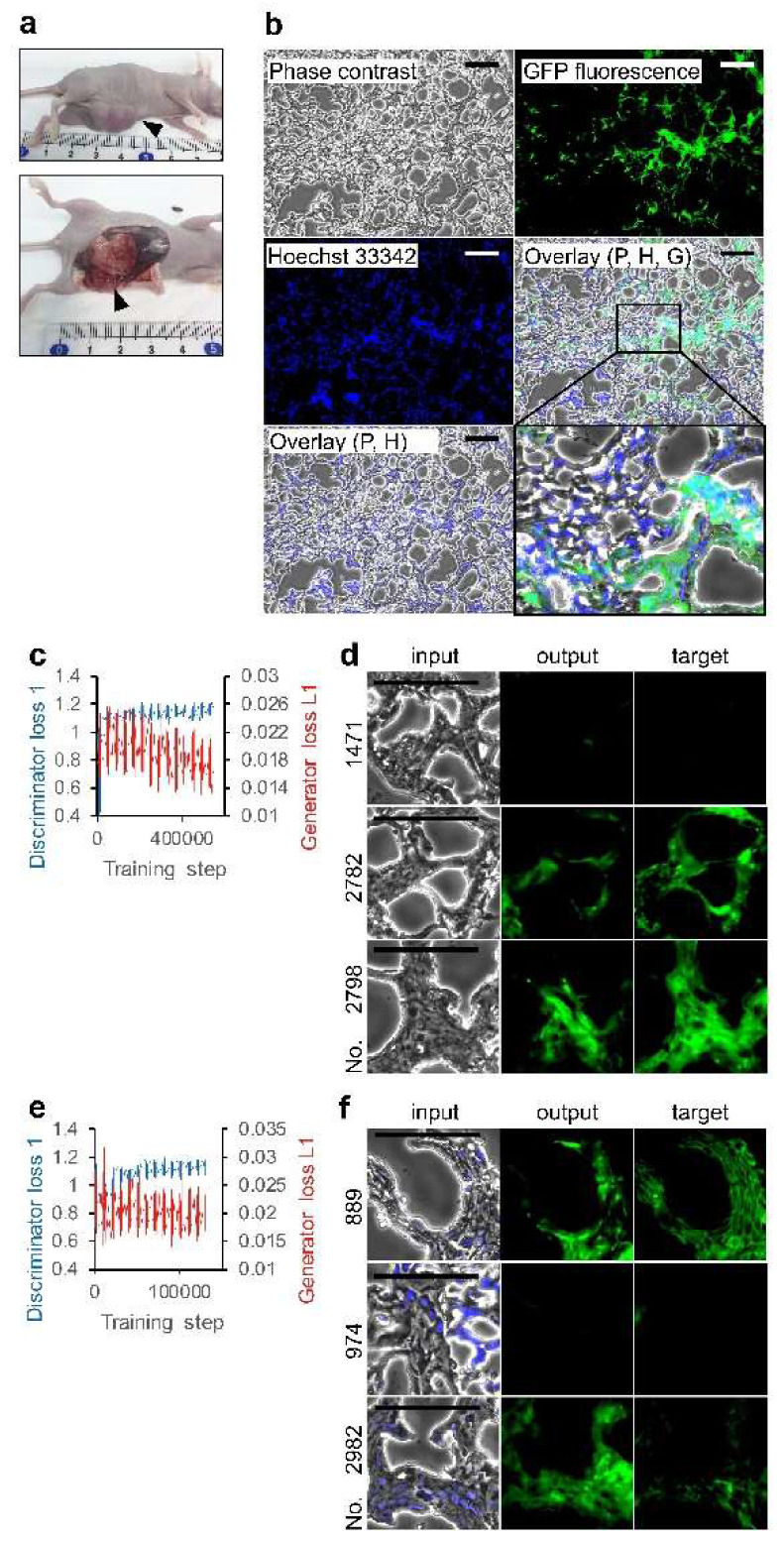
Deep-learning workflows of microscopic image analysis are sufficient for handling the contextual variations because they employ biological samples and have numerous tasks. The use of well-defined annotated images is important for the workflow. Cancer stem cells (CSCs) are identified by specific cell markers. These CSCs were extensively characterized by the stem cell (SC)-like gene expression and proliferation mechanisms for the development of tumors. In contrast, the morphological characterization remains elusive. This study aims to investigate the segmentation of CSCs in phase contrast imaging using conditional generative adversarial networks (CGAN). Artificial intelligence (AI) was trained using fluorescence images of the -Green fluorescence protein, the expression of which was maintained in CSCs, and the phase contrast images. The AI model segmented the CSC region in the phase contrast image of the CSC cultures and tumor model. By selecting images for training, several values for measuring segmentation quality increased. Moreover, nucleus fluorescence overlaid-phase contrast was effective for increasing the values. We show the possibility of mapping CSC morphology to the condition of undifferentiation using deep-learning CGAN workflows.
深度学习的微观图像分析工作流程足以处理上下文变化,因为它们使用生物样本并具有许多任务。使用定义明确的注释图像对于工作流程很重要。癌症干细胞 (CSC) 通过特定的细胞标记物来识别。这些 CSC 通过类似于干细胞 (SC) 的基因表达和增殖机制来广泛表征肿瘤的发展。相比之下,形态特征仍然难以捉摸。本研究旨在使用条件生成对抗网络 (CGAN) 研究相位对比成像中的 CSC 分割。使用表达于 CSC 中的绿色荧光蛋白的荧光图像和相位对比图像对人工智能 (AI) 进行训练。AI 模型对 CSC 培养物和肿瘤模型的相位对比图像中的 CSC 区域进行分割。通过选择用于训练的图像,用于测量分割质量的几个值增加了。此外,细胞核荧光叠加相位对比对于增加这些值是有效的。我们展示了使用深度学习 CGAN 工作流程将 CSC 形态映射到未分化状态的可能性。