Shen Guiquan, Wang Rujia, Gao Bo, Zhang Zhongwen, Wu Guipeng, Pope Whitney
Affiliated Hospital of Guizhou Medical University, Guiyang, China.
Tangshan Gongren Hospital, Tangshan, China.
Front Oncol. 2020 Jun 2;10:852. doi: 10.3389/fonc.2020.00852. eCollection 2020.
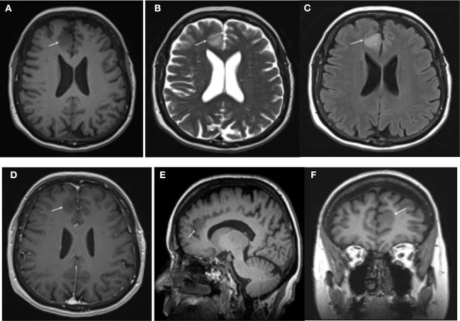
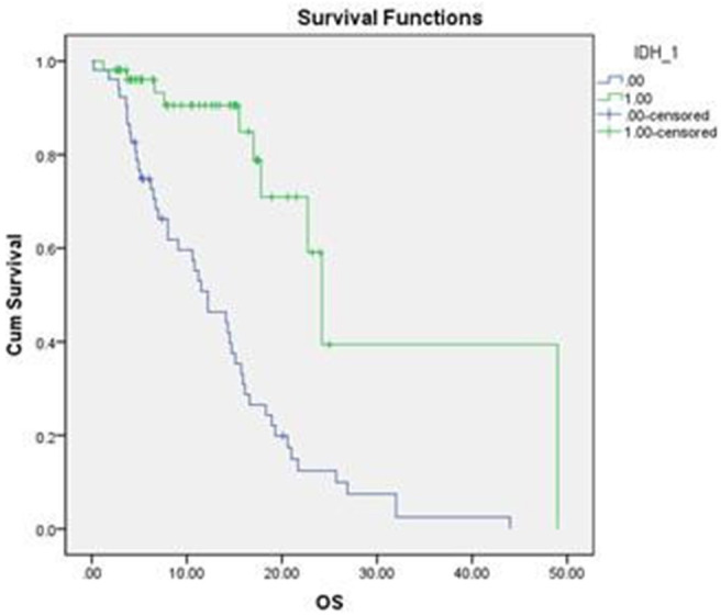
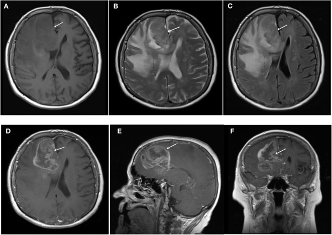
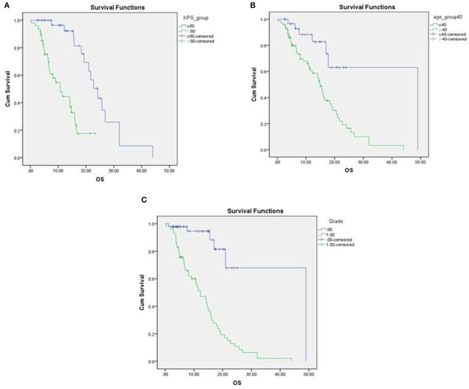
To investigate the associations of MRI radiological features and prognosis of glioma with the status of isocitrate dehydrogenase 1 (IDH1). A total of 116 patients with gliomas were retrospectively recruited from January 2013 to December 2015. All patients were undergone routine MRI (T1WI, T2WI, T2-FLAIR) scanning and contrast-enhanced MRI T1WI before surgery. The following imaging features were included: tumor location, diameter, the pattern of growth, boundary, the degree of enhancement, mass effect, edema, cross the middle line, under the ependyma. χ and Fisher's exact probability tests were used to determine the significance of associations between MRI features and IDH1 mutation of glioma. The survival distributions were estimated using Kaplan-Meier compared by Log-rank test. Univariate and multivariate analyses were performed using Cox regression. Gliomas with IDH1 mutant were significantly more likely to exhibit homogeneous signal intensity ( = 0.009) on non-contrast MRI protocols and less contrast enhancement ( = 0.000) on contrast enhanced T1WI. IDH1 mutant type glioma was more inclined to cross the midline to invade contralateral hemisphere ( = 0.001). The overall survival between IDH1 mutated and wild type glioma were significantly different ( = 0.000), age ≤ 40 ( = 0.003), KPS scores > 80 before operation ( = 0.000) and low grade glioma ( = 0.000). Our results suggest IDH1 mutant in gliomas is more likely to exhibit homogeneous signal intensity, less contrast enhancement and more inclined to cross the midline. Patients with IDH1 mutated, age ≤ 40, KPS scores > 80 before operation and low-grade glioma may have a longer life and better prognosis.
探讨异柠檬酸脱氢酶1(IDH1)状态与胶质瘤MRI影像学特征及预后的相关性。回顾性纳入2013年1月至2015年12月期间的116例胶质瘤患者。所有患者在手术前行常规MRI(T1WI、T2WI、T2-FLAIR)扫描及增强MRI T1WI扫描。纳入的影像学特征包括:肿瘤位置、直径、生长方式、边界、强化程度、占位效应、水肿、是否跨越中线、是否位于室管膜下。采用χ检验和Fisher精确概率检验确定MRI特征与胶质瘤IDH1突变之间关联的显著性。采用Kaplan-Meier法估计生存分布,并通过Log-rank检验进行比较。使用Cox回归进行单因素和多因素分析。IDH1突变型胶质瘤在非增强MRI序列上更易表现为均匀信号强度(P = 0.009),在增强T1WI上强化程度更低(P = 0.000)。IDH1突变型胶质瘤更倾向于跨越中线侵犯对侧半球(P = 0.001)。IDH1突变型和野生型胶质瘤的总生存期存在显著差异(P = 0.000),年龄≤40岁(P = 0.003)、术前KPS评分>80分(P = 0.000)以及低级别胶质瘤(P = 0.000)。我们的结果表明,胶质瘤中的IDH1突变更易表现为均匀信号强度、强化程度更低且更倾向于跨越中线。IDH1突变、年龄≤40岁、术前KPS评分>80分以及低级别胶质瘤患者可能具有更长的生存期和更好的预后。