• 文献检索
  • 文档翻译
  • 深度研究
  • 学术资讯
  • Suppr Zotero 插件Zotero 插件
  • 邀请有礼
  • 套餐&价格
  • 历史记录
应用&插件
Suppr Zotero 插件Zotero 插件浏览器插件Mac 客户端Windows 客户端微信小程序
定价
高级版会员购买积分包购买API积分包
服务
文献检索文档翻译深度研究API 文档MCP 服务
关于我们
关于 Suppr公司介绍联系我们用户协议隐私条款
关注我们

Suppr 超能文献

核心技术专利:CN118964589B侵权必究
粤ICP备2023148730 号-1Suppr @ 2026

文献检索

告别复杂PubMed语法,用中文像聊天一样搜索,搜遍4000万医学文献。AI智能推荐,让科研检索更轻松。

立即免费搜索

文件翻译

保留排版,准确专业,支持PDF/Word/PPT等文件格式,支持 12+语言互译。

免费翻译文档

深度研究

AI帮你快速写综述,25分钟生成高质量综述,智能提取关键信息,辅助科研写作。

立即免费体验

三维建模与打印技术对经皮骶骨螺钉内固定术的手术成功率有影响吗?

Do three-dimensional modeling and printing technologies have an impact on the surgical success of percutaneous transsacral screw fixation?

作者信息

Durusoy Serhat, Akdoğan Volkan, Paksoy Ahmet Emre

机构信息

Yozgat Bozok Üniversitesi Tıp Fakültesi Eğitim ve Araştırma Hastanesi Ortopedi ve Travmatoloji Kliniği, 66900 Yozgat, Türkiye.

出版信息

Jt Dis Relat Surg. 2020;31(2):273-280. doi: 10.5606/ehc.2020.73115. Epub 2020 Jun 18.

DOI:10.5606/ehc.2020.73115
PMID:32584725
原文链接:https://pmc.ncbi.nlm.nih.gov/articles/PMC7489170/
Abstract

OBJECTIVES

This study aims to determine the role of computed tomography (CT)-derived templates, produced by three- dimensional (3D) modeling, image processing and printing technology, in percutaneous transsacral screw fixation and evaluate the effects of their use on surgical success.

MATERIALS AND METHODS

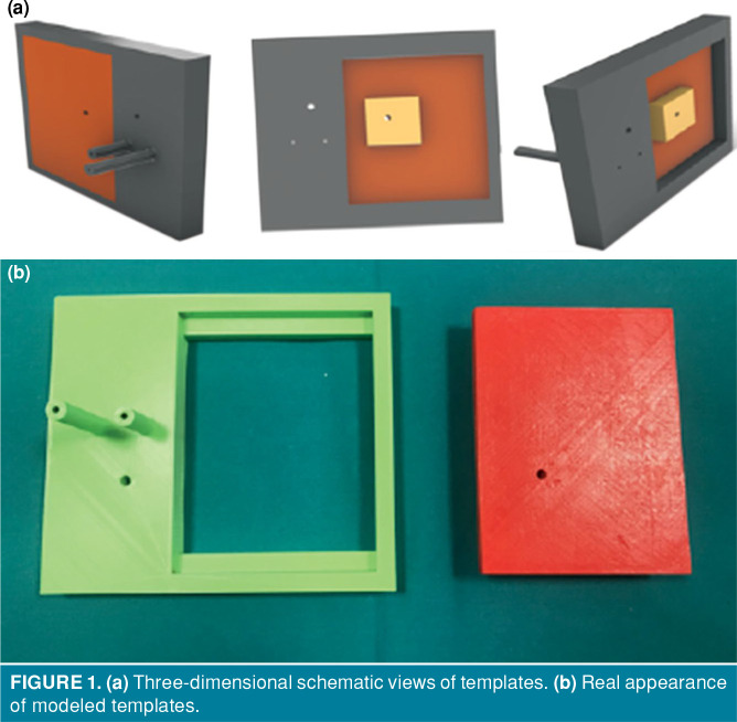
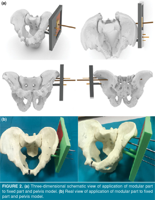
This prospective study conducted between June 2018 and December 2019 utilized 15 composite pelvis models for transsacral-transiliac screw fixation. For the procedure, modeled templates were utilized for wiring on the left side of the pelvis models, while the conventional method was performed on the right side of the pelvis models. In the computed tomography images acquired after wiring, appropriate wire position was evaluated.

RESULTS

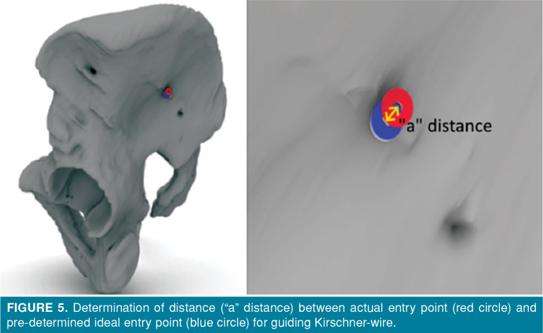
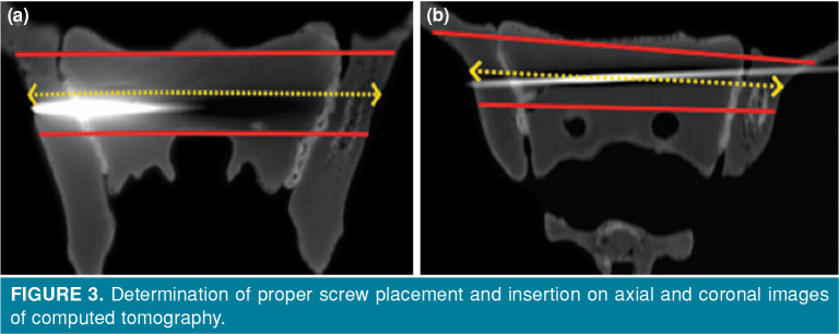
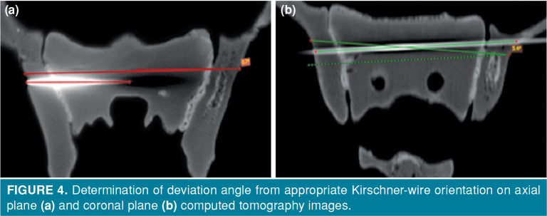
The placed wires held the S1 body appropriately in all of the procedures with or without template use. With the template use, the wires were placed appropriately in the surgical bone corridor suitable for the transsacral-transiliac screw fixation in all of the models. However, with the conventional methods, the wires were not placed in the safe surgical bone corridor in four models. The wire deviation angle in the axial plane was significantly lower in the template group (p=0.001), whereas it was not different between the template group and the conventional method group in the coronal plane (p=0.054). The amount of deviation from the ideal wire entry site was significantly reduced in the template group compared to the conventional method group (p=0.001).

CONCLUSION

With the use of 3D modeling and printing technology, CT-derived templates can be produced and utilized for transsacral screw fixation procedures and their use increases surgical success by reducing the surgical margin of error.

摘要

目的

本研究旨在确定通过三维(3D)建模、图像处理和打印技术生成的计算机断层扫描(CT)衍生模板在经皮骶骨螺钉固定中的作用,并评估其使用对手术成功率的影响。

材料与方法

本前瞻性研究于2018年6月至2019年12月进行,使用15个复合骨盆模型进行骶骨-髂骨螺钉固定。在手术过程中,将建模模板用于骨盆模型左侧的布线,而右侧骨盆模型采用传统方法。在布线后获取的计算机断层扫描图像中,评估导线的合适位置。

结果

在所有使用或未使用模板的手术中,放置的导线均能妥善固定S1椎体。使用模板时,在所有模型中,导线均被正确放置在适合骶骨-髂骨螺钉固定的手术骨通道内。然而,采用传统方法时,有4个模型的导线未放置在安全的手术骨通道内。模板组在轴向平面的导线偏差角度显著更低(p = 0.001),而在冠状平面,模板组与传统方法组之间无差异(p = 0.054)。与传统方法组相比,模板组从理想导线进入点的偏差量显著减少(p = 0.001)。

结论

通过使用3D建模和打印技术,可以制作CT衍生模板并将其用于骶骨螺钉固定手术,其使用通过减少手术误差范围提高了手术成功率。

https://cdn.ncbi.nlm.nih.gov/pmc/blobs/907b/7489170/9e7bc3301d1a/JDRS-2020-31-2-273-280-F5.jpg
https://cdn.ncbi.nlm.nih.gov/pmc/blobs/907b/7489170/36211fe61e8a/JDRS-2020-31-2-273-280-F1.jpg
https://cdn.ncbi.nlm.nih.gov/pmc/blobs/907b/7489170/b123522e40ce/JDRS-2020-31-2-273-280-F2.jpg
https://cdn.ncbi.nlm.nih.gov/pmc/blobs/907b/7489170/8ccacdaf61ac/JDRS-2020-31-2-273-280-F3.jpg
https://cdn.ncbi.nlm.nih.gov/pmc/blobs/907b/7489170/25ccdc294f40/JDRS-2020-31-2-273-280-F4.jpg
https://cdn.ncbi.nlm.nih.gov/pmc/blobs/907b/7489170/9e7bc3301d1a/JDRS-2020-31-2-273-280-F5.jpg

相似文献

1
Do three-dimensional modeling and printing technologies have an impact on the surgical success of percutaneous transsacral screw fixation?三维建模与打印技术对经皮骶骨螺钉内固定术的手术成功率有影响吗?
Jt Dis Relat Surg. 2020;31(2):273-280. doi: 10.5606/ehc.2020.73115. Epub 2020 Jun 18.
2
Transsacral Osseous Corridor Anatomy Is More Amenable To Screw Insertion In Males: A Biomorphometric Analysis of 280 Pelves.经骶骨骨性通道解剖结构更有利于男性进行螺钉置入:对280例骨盆的生物形态测量分析
Clin Orthop Relat Res. 2016 Oct;474(10):2304-11. doi: 10.1007/s11999-016-4954-5. Epub 2016 Jul 8.
3
Factors influencing the accuracy of iliosacral screw insertion using 3D fluoroscopic navigation.使用三维荧光透视导航影响髂骶螺钉置入准确性的因素。
Arch Orthop Trauma Surg. 2019 Feb;139(2):189-195. doi: 10.1007/s00402-018-3055-1. Epub 2018 Oct 29.
4
[Implantation of iliosacral screws. Simulation of optimal placement by 3-dimensional X-ray computed tomography].[髂骶螺钉植入。通过三维X线计算机断层扫描模拟最佳置入位置]
Rev Chir Orthop Reparatrice Appar Mot. 2000 Jun;86(4):360-9.
5
Biomechanical study of transsacral-transiliac screw fixation versus lumbopelvic fixation and bilateral triangular fixation for "H"- and "U"-type sacrum fractures with traumatic spondylopelvic dissociation: a finite element analysis study.经关节突-髂骨螺钉固定与腰骶骨盆固定和双侧三角固定治疗创伤性腰骶骨盆分离伴“H”型和“U”型骶骨骨折的生物力学研究:有限元分析研究。
J Orthop Surg Res. 2021 Jul 3;16(1):428. doi: 10.1186/s13018-021-02581-5.
6
Gender-Associated Differences in Sacral Morphology Do Not Affect Feasibility Rates of Transsacral Screw Insertion. Radioanatomic Investigation Based on Pelvic Cross-sectional Imaging of 200 Individuals.性别相关的骶骨形态差异不会影响经骶骨螺钉插入的可行性。基于 200 例个体骨盆横断面成像的影像学研究。
Spine (Phila Pa 1976). 2020 Apr 1;45(7):421-430. doi: 10.1097/BRS.0000000000003293.
7
Biomechanical comparison of standard iliosacral screw fixation to transsacral locked screw fixation in a type C zone II pelvic fracture model.生物力学比较标准的髂骶螺钉固定与经骶骨锁定螺钉固定在 C 型骨盆骨折模型中的 II 区。
J Orthop Trauma. 2013 Sep;27(9):521-6. doi: 10.1097/BOT.0b013e3182781102.
8
A useful intraoperative technique for transiliac-transsacral screws: a point-to-point coaxial guide apparatus.一种用于经髂-经骶螺钉的有用术中技术:点对点同轴导向装置。
J Orthop Surg Res. 2021 Jan 28;16(1):89. doi: 10.1186/s13018-021-02239-2.
9
Quantification of the Safe Zone of the First to Third Sacral Segments for Transiliac-Transsacral Screw Fixation in Normal and Dysmorphic Sacra.正常和畸形骶骨经髂骨-骶骨螺钉固定的第一至第三骶段安全区的定量评估。
Orthopedics. 2024 Jan-Feb;47(1):e13-e18. doi: 10.3928/01477447-20230531-06. Epub 2023 Jun 5.
10
Assessment of osseous corridor for transiliac-transsacral screws and clinical applications: Computational simulation study.评估经髂骨-经骶骨螺钉的骨道及其临床应用:计算模拟研究。
Orthop Traumatol Surg Res. 2020 Sep;106(5):863-867. doi: 10.1016/j.otsr.2020.03.023. Epub 2020 Jun 18.

引用本文的文献

1
Evaluation of the true lateral fluoroscopic projection for the relation of the S1 recess/foramen to safe corridors in transiliac-transsacral screw placement in human cadaveric pelves.评价 S1 隐窝/孔与经髂骨-骶骨螺钉放置在人体骨盆标本中的安全通道之间关系的真正侧位透视投影。
Eur J Orthop Surg Traumatol. 2024 Nov 28;35(1):31. doi: 10.1007/s00590-024-04157-5.
2
Application of 3D printed models in the surgical treatment of spinal deformity.3D打印模型在脊柱畸形外科治疗中的应用。
Am J Transl Res. 2022 Sep 15;14(9):6341-6348. eCollection 2022.
3
Does a Customized 3D Printing Plate Based on Virtual Reduction Facilitate the Restoration of Original Anatomy in Fractures?

本文引用的文献

1
Is pelvic mapping applicable in iliosacral screw fixation to determine screw entry point and screw trajectory?骨盆测绘在髂骶螺钉固定中是否适用于确定螺钉进钉点和螺钉轨迹?
Eklem Hastalik Cerrahisi. 2019 Dec;30(3):252-8. doi: 10.5606/ehc.2019.66489.
2
Comparative study of sacroiliac screw placement guided by 3D-printed template technology and X-ray fluoroscopy.三维打印模板技术与 X 射线透视引导下骶髂螺钉置钉的对比研究。
Arch Orthop Trauma Surg. 2020 Jan;140(1):11-17. doi: 10.1007/s00402-019-03207-6. Epub 2019 May 24.
3
Rate of intraoperative problems during sacroiliac screw removal: expect the unexpected.
基于虚拟复位的定制3D打印接骨板是否有助于骨折后原始解剖结构的恢复?
J Pers Med. 2022 Jun 2;12(6):927. doi: 10.3390/jpm12060927.
4
A Systematic Review and Meta-Analysis of 3D Printing Technology for the Treatment of Acetabular Fractures.3D 打印技术治疗髋臼骨折的系统评价和荟萃分析。
Biomed Res Int. 2021 Aug 17;2021:5018791. doi: 10.1155/2021/5018791. eCollection 2021.
5
Is SARS-CoV-2 Infection a Risk Factor for Early Pregnancy Loss? ACE2 and TMPRSS2 Coexpression and Persistent Replicative Infection in Primitive Trophoblast.SARS-CoV-2 感染是否是早期妊娠丢失的一个危险因素?ACE2 和 TMPRSS2 的共表达与原始滋养细胞中的持续复制性感染。
J Infect Dis. 2021 Dec 8;224(Suppl 6):S660-S669. doi: 10.1093/infdis/jiab309.
骶髂螺钉取出术中出现问题的发生率:做好应对意外情况的准备。
BMC Surg. 2019 Apr 15;19(1):39. doi: 10.1186/s12893-019-0501-0.
4
Application of the Guiding Template Designed by Three-dimensional Printing Data for the Insertion of Sacroiliac Screws: a New Clinical Technique.应用三维打印数据设计的导板置入骶髂螺钉:一种新的临床技术。
Curr Med Sci. 2018 Dec;38(6):1090-1095. doi: 10.1007/s11596-018-1988-9. Epub 2018 Dec 7.
5
A novel patient-specific three-dimensional-printed external template to guide iliosacral screw insertion: a retrospective study.一种用于引导骶髂螺钉植入的新型患者特异性三维打印外部模板:一项回顾性研究。
BMC Musculoskelet Disord. 2018 Nov 13;19(1):397. doi: 10.1186/s12891-018-2320-3.
6
Intraoperative CT and Surgical Navigation for Iliosacral Screws: Technique for Patients With Sacral Dysmorphism.术中 CT 和手术导航在骶髂螺钉中的应用:骶骨发育不良患者的技术。
J Orthop Trauma. 2018 Aug;32 Suppl 1:S24-S25. doi: 10.1097/BOT.0000000000001213.
7
Epidemiology of Pelvic Ring Fractures in a Level 1 Trauma Center in the Netherlands.荷兰一家一级创伤中心骨盆环骨折的流行病学研究
Hip Pelvis. 2017 Dec;29(4):253-261. doi: 10.5371/hp.2017.29.4.253. Epub 2017 Dec 1.
8
A comparison study of pelvic fractures and associated abdominal injuries between pediatric and adult blunt trauma patients.小儿与成人钝性创伤患者骨盆骨折及相关腹部损伤的比较研究。
J Pediatr Surg. 2017 Mar;52(3):386-389. doi: 10.1016/j.jpedsurg.2016.09.055. Epub 2016 Sep 23.
9
A Useful Preoperative Planning Technique for Transiliac-Transsacral Screws.一种用于髂骨-骶骨螺钉的有用术前规划技术。
J Orthop Trauma. 2017 Jan;31(1):e25-e31. doi: 10.1097/BOT.0000000000000708.
10
2D versus 3D fluoroscopy-based navigation in posterior pelvic fixation: review of the literature on current technology.二维与三维透视导航在后盆骨固定术中的应用:当前技术文献综述。
Int J Comput Assist Radiol Surg. 2017 Jan;12(1):69-76. doi: 10.1007/s11548-016-1465-5. Epub 2016 Aug 8.