• 文献检索
  • 文档翻译
  • 深度研究
  • 学术资讯
  • Suppr Zotero 插件Zotero 插件
  • 邀请有礼
  • 套餐&价格
  • 历史记录
应用&插件
Suppr Zotero 插件Zotero 插件浏览器插件Mac 客户端Windows 客户端微信小程序
定价
高级版会员购买积分包购买API积分包
服务
文献检索文档翻译深度研究API 文档MCP 服务
关于我们
关于 Suppr公司介绍联系我们用户协议隐私条款
关注我们

Suppr 超能文献

核心技术专利:CN118964589B侵权必究
粤ICP备2023148730 号-1Suppr @ 2026

文献检索

告别复杂PubMed语法,用中文像聊天一样搜索,搜遍4000万医学文献。AI智能推荐,让科研检索更轻松。

立即免费搜索

文件翻译

保留排版,准确专业,支持PDF/Word/PPT等文件格式,支持 12+语言互译。

免费翻译文档

深度研究

AI帮你快速写综述,25分钟生成高质量综述,智能提取关键信息,辅助科研写作。

立即免费体验

女性患者正畸治疗中咀嚼肌及周围软组织厚度变化的回顾性研究。

Thickness change of masseter muscles and the surrounding soft tissues in female patients during orthodontic treatment: a retrospective study.

机构信息

Department of Orthodontics, Peking University School and Hospital of Stomatology, 22 Zhongguancun Avenue South, Haidian District, Beijing, 100081, PR China.

National Engineering Laboratory for Digital and Material Technology of Stomatology, 22 Zhongguancun Avenue South, Haidian District, Beijing, 100081, PR China.

出版信息

BMC Oral Health. 2020 Jun 29;20(1):181. doi: 10.1186/s12903-020-01168-6.

DOI:10.1186/s12903-020-01168-6
PMID:32600308
原文链接:https://pmc.ncbi.nlm.nih.gov/articles/PMC7325017/
Abstract

BACKGROUND

Facial esthetics is a major concern of orthodontic patients. This study aims to evaluate orthodontic treatment-related thickness changes of the masseter muscles and surrounding soft tissues and the potential factors that would influence these changes during orthodontic treatment in female adults.

METHODS

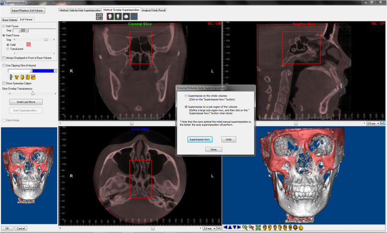
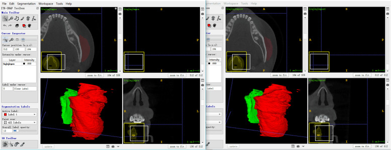
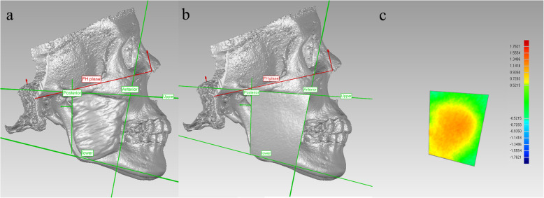
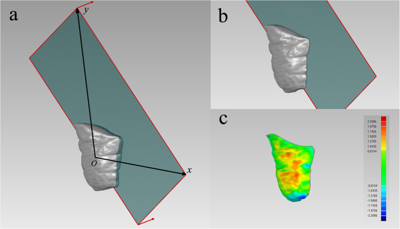
Forty-two female adult patients were included in this retrospective study and were divided into extraction (n = 22) and nonextraction (n = 20) groups. Pretreatment and posttreatment cone-beam computed tomography (CBCT) images were superimposed and reconstructed. The thickness changes of the masseter area of facial soft tissue (MAS), masseter muscles (MM) and surrounding fat tissue (FT) were measured. Pretreatment age, treatment duration, sagittal relationship (ANB), and vertical relationship (Frankfort-mandibular plane angle, FMA)-related MAS, MM and FT changes were compared between extraction and nonextraction groups. Spearman's correlation coefficient was calculated between the above variables. Regression analysis was conducted to confirm the causal relations of the variables.

RESULTS

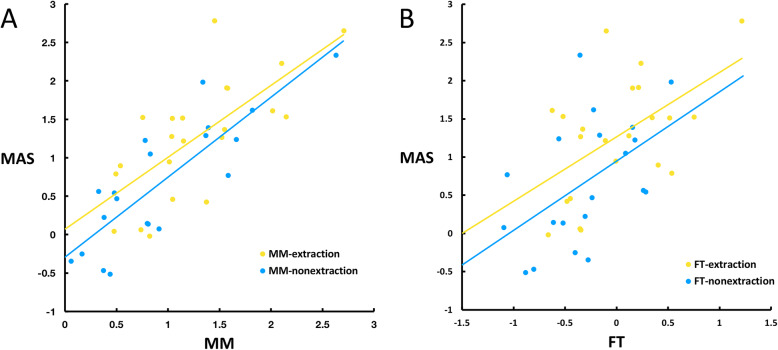
The thickness of MAS and MM significantly decreased in both groups, with larger decreases (> 1 mm) in the extraction group. There were strong correlations (r > 0.7) between the thickness decrease in MAS and MM in both groups and moderate correlations (r > 0.4) between MAS and FT in the nonextraction group. A significantly greater decrease of MAS and MM were found to be moderately correlated with a smaller FMA (r > 0.4) in the extraction group. Scatter plots and regression analysis confirmed these correlations.

CONCLUSIONS

Masseter muscles and the surrounding soft tissue exhibited a significant decrease in thickness during orthodontic treatment in female adults. Low-angle patients experienced a greater decrease in soft tissue thickness in the masseter area in the extraction case. But the thickness changes were clinically very small in most patients.

摘要

背景

面部美学是正畸患者关注的主要问题。本研究旨在评估女性成年人正畸治疗相关的咀嚼肌及周围软组织厚度变化,并探讨正畸治疗过程中可能影响这些变化的因素。

方法

本回顾性研究纳入 42 名女性成年患者,分为拔牙组(n=22)和非拔牙组(n=20)。对治疗前后锥形束 CT(CBCT)图像进行叠加和重建,测量面部软组织(MAS)、咀嚼肌(MM)和周围脂肪组织(FT)的咀嚼肌区厚度变化。比较拔牙组和非拔牙组治疗前年龄、治疗时间、矢状关系(ANB)和垂直关系(法兰克福-下颌平面角,FMA)与 MAS、MM 和 FT 变化的相关性。采用 Spearman 相关系数分析上述变量之间的相关性。进行回归分析以确认变量之间的因果关系。

结果

两组 MAS 和 MM 的厚度均显著减小,拔牙组减小幅度更大(>1mm)。两组 MAS 和 MM 的厚度减小之间存在很强的相关性(r>0.7),非拔牙组 MAS 和 FT 之间存在中度相关性(r>0.4)。在拔牙组中,MAS 和 MM 的显著减小与较小的 FMA 呈中度相关(r>0.4)。散点图和回归分析证实了这些相关性。

结论

女性成年人在正畸治疗过程中咀嚼肌及周围软组织厚度显著减小。低角患者在拔牙病例中咀嚼肌区软组织厚度减小较大。但在大多数患者中,厚度变化在临床上非常小。

https://cdn.ncbi.nlm.nih.gov/pmc/blobs/dda3/7325017/2da16c87d9b5/12903_2020_1168_Fig5_HTML.jpg
https://cdn.ncbi.nlm.nih.gov/pmc/blobs/dda3/7325017/8b460eca3857/12903_2020_1168_Fig1_HTML.jpg
https://cdn.ncbi.nlm.nih.gov/pmc/blobs/dda3/7325017/8b42d2f839be/12903_2020_1168_Fig2_HTML.jpg
https://cdn.ncbi.nlm.nih.gov/pmc/blobs/dda3/7325017/6c530cbe0b67/12903_2020_1168_Fig3_HTML.jpg
https://cdn.ncbi.nlm.nih.gov/pmc/blobs/dda3/7325017/c7e7ac949e79/12903_2020_1168_Fig4_HTML.jpg
https://cdn.ncbi.nlm.nih.gov/pmc/blobs/dda3/7325017/2da16c87d9b5/12903_2020_1168_Fig5_HTML.jpg

相似文献

1
Thickness change of masseter muscles and the surrounding soft tissues in female patients during orthodontic treatment: a retrospective study.女性患者正畸治疗中咀嚼肌及周围软组织厚度变化的回顾性研究。
BMC Oral Health. 2020 Jun 29;20(1):181. doi: 10.1186/s12903-020-01168-6.
2
Evaluation of masseter muscle morphology in different types of malocclusions using cone beam computed tomography.使用锥形束计算机断层扫描评估不同类型错牙合畸形中的咬肌形态。
Int Orthod. 2014 Mar;12(1):32-48. doi: 10.1016/j.ortho.2013.12.003. Epub 2014 Jan 21.
3
Relationship between masseter muscle thickness and maxillofacial morphology in pre-orthodontic treatment patients.正畸治疗前患者咬肌厚度与颌面形态的关系。
Int Orthod. 2018 Dec;16(4):698-711. doi: 10.1016/j.ortho.2018.09.015. Epub 2018 Oct 19.
4
Evaluation of masseter muscles in relation to treatment with removable bite-blocks in dolichofacial growing subjects: A prospective controlled study.长面型生长发育期受试者使用可摘式咬垫治疗时咬肌的评估:一项前瞻性对照研究。
Am J Orthod Dentofacial Orthop. 2017 Jun;151(6):1058-1064. doi: 10.1016/j.ajodo.2016.10.035.
5
Three-dimensional evaluation of masseter muscle in different vertical facial patterns: a cross-sectional study in growing children.不同垂直面型生长儿童中咀嚼肌的三维评估:一项横断面研究。
Ultrason Imaging. 2013 Oct;35(4):307-17. doi: 10.1177/0161734613502468.
6
Masseter muscle thickness in different skeletal morphology: an ultrasonographic study.不同骨骼形态下的咬肌厚度:一项超声研究
Indian J Dent Res. 2010 Jul-Sep;21(3):402-7. doi: 10.4103/0970-9290.70812.
7
Validity and reliability of masseter muscles segmentation from the transverse sections of Cone-Beam CT scans compared with MRI scans.比较锥形束 CT 扫描的横断面与 MRI 扫描对咀嚼肌的分割的有效性和可靠性。
Int J Comput Assist Radiol Surg. 2022 Apr;17(4):751-759. doi: 10.1007/s11548-021-02513-y. Epub 2021 Oct 8.
8
Facial Soft Tissue Characteristics Among Sagittal and Vertical Skeletal Patterns: A Cone-Beam Computed Tomography Study.矢状面和垂直面骨骼类型中的面部软组织特征:一项锥形束计算机断层扫描研究
Cureus. 2023 Aug 31;15(8):e44428. doi: 10.7759/cureus.44428. eCollection 2023 Aug.
9
Comparative data on facial morphology and muscle thickness using ultrasonography.使用超声检查对面部形态和肌肉厚度的比较数据。
Eur J Orthod. 2005 Dec;27(6):562-7. doi: 10.1093/ejo/cji052. Epub 2005 Aug 31.
10
Masseter muscle thickness and mechanical advantage in relation to vertical craniofacial morphology in children.儿童咬肌厚度及机械优势与垂直颅面形态的关系
Acta Odontol Scand. 2008 Feb;66(1):23-30. doi: 10.1080/00016350701884604.

引用本文的文献

1
Anatomic and functional masseter muscle adaptation following orthognathic surgery-MRI analysis in 3 years of follow-up.正颌外科手术后咬肌的解剖学和功能适应性——3年随访的MRI分析
Maxillofac Plast Reconstr Surg. 2024 Jul 19;46(1):26. doi: 10.1186/s40902-024-00437-6.
2
Automatic Masseter Muscle Accurate Segmentation from CBCT Using Deep Learning-Based Model.基于深度学习模型从锥形束计算机断层扫描(CBCT)中自动精确分割咬肌
J Clin Med. 2022 Dec 21;12(1):55. doi: 10.3390/jcm12010055.
3
Validity and reliability of masseter muscles segmentation from the transverse sections of Cone-Beam CT scans compared with MRI scans.

本文引用的文献

1
Cranial Base Superimposition of Cone-Beam Computed Tomography Images: A Voxel-Based Protocol Validation.锥形束计算机断层扫描图像的颅底叠加:基于体素的方案验证
J Craniofac Surg. 2019 Sep;30(6):1809-1814. doi: 10.1097/SCS.0000000000005503.
2
Changes in buccal facial depth of female patients after extraction and nonextraction orthodontic treatments: A preliminary study.拔牙与非拔牙正畸治疗后女性患者颊面部深度的变化:一项初步研究。
Korean J Orthod. 2018 May;48(3):172-181. doi: 10.4041/kjod.2018.48.3.172. Epub 2018 Apr 20.
3
Repeated injections of botulinum toxin into the masseter muscle induce bony changes in human adults: A longitudinal study.
比较锥形束 CT 扫描的横断面与 MRI 扫描对咀嚼肌的分割的有效性和可靠性。
Int J Comput Assist Radiol Surg. 2022 Apr;17(4):751-759. doi: 10.1007/s11548-021-02513-y. Epub 2021 Oct 8.
重复向咬肌注射肉毒杆菌毒素可导致成年人体骨骼变化:一项纵向研究。
Korean J Orthod. 2017 Jul;47(4):222-228. doi: 10.4041/kjod.2017.47.4.222. Epub 2017 May 26.
4
Changes in occlusal function after extraction of premolars: 2-year follow-up.磨牙拔除后咬合功能的变化:2 年随访。
Angle Orthod. 2017 Sep;87(5):703-708. doi: 10.2319/112116-836.1. Epub 2017 May 9.
5
Three-dimensional computed tomographic assessment of mandibular muscles in growing subjects with different vertical facial patterns.对具有不同垂直面部模式的生长发育期受试者下颌肌肉的三维计算机断层扫描评估
Aust Orthod J. 2016 May;32(1):2-17.
6
Performance of cone-beam computed tomography and multidetector computed tomography in diagnostic imaging of the midface: A comparative study on Phantom and cadaver head scans.锥形束计算机断层扫描和多排螺旋计算机断层扫描在中面部诊断成像中的性能:对 Phantom 和尸体头部扫描的比较研究。
Eur Radiol. 2017 Feb;27(2):790-800. doi: 10.1007/s00330-016-4387-2. Epub 2016 May 11.
7
Effect of different incisor movements on the soft tissue profile measured in reference to a rough-surfaced palatal implant.以粗糙表面的腭部种植体为参照测量不同切牙移动对软组织轮廓的影响。
Am J Orthod Dentofacial Orthop. 2016 Mar;149(3):349-57. doi: 10.1016/j.ajodo.2015.08.017.
8
Soft-tissue changes in Class II malocclusion patients treated with extractions: a systematic review.拔牙治疗的安氏II类错颌患者的软组织变化:一项系统评价
Eur J Orthod. 2016 Dec;38(6):631-637. doi: 10.1093/ejo/cjv083. Epub 2015 Nov 29.
9
Assessment of image quality in soft tissue and bone visualization tasks for a dedicated extremity cone-beam CT system.专用四肢锥形束CT系统在软组织和骨骼可视化任务中的图像质量评估。
Eur Radiol. 2015 Jun;25(6):1742-51. doi: 10.1007/s00330-014-3546-6. Epub 2015 Jan 20.
10
Three-dimensional imaging methods for quantitative analysis of facial soft tissues and skeletal morphology in patients with orofacial clefts: a systematic review.口腔颌面裂患者面部软组织和骨骼形态定量分析的三维成像方法:一项系统评价
PLoS One. 2014 Apr 7;9(4):e93442. doi: 10.1371/journal.pone.0093442. eCollection 2014.