• 文献检索
  • 文档翻译
  • 深度研究
  • 学术资讯
  • Suppr Zotero 插件Zotero 插件
  • 邀请有礼
  • 套餐&价格
  • 历史记录
应用&插件
Suppr Zotero 插件Zotero 插件浏览器插件Mac 客户端Windows 客户端微信小程序
定价
高级版会员购买积分包购买API积分包
服务
文献检索文档翻译深度研究API 文档MCP 服务
关于我们
关于 Suppr公司介绍联系我们用户协议隐私条款
关注我们

Suppr 超能文献

核心技术专利:CN118964589B侵权必究
粤ICP备2023148730 号-1Suppr @ 2026

文献检索

告别复杂PubMed语法,用中文像聊天一样搜索,搜遍4000万医学文献。AI智能推荐,让科研检索更轻松。

立即免费搜索

文件翻译

保留排版,准确专业,支持PDF/Word/PPT等文件格式,支持 12+语言互译。

免费翻译文档

深度研究

AI帮你快速写综述,25分钟生成高质量综述,智能提取关键信息,辅助科研写作。

立即免费体验

通过抑制衰老,激活的 p38 MAPKα 和 mTORC1 的共抑制增强了 SR1 扩增的人脐血 CD34 细胞中 HSCs 的干性维持。

Coinhibition of activated p38 MAPKα and mTORC1 potentiates stemness maintenance of HSCs from SR1-expanded human cord blood CD34 cells via inhibition of senescence.

机构信息

Center for Translational Medicine, Union Hospital, Tongji Medical College, Huazhong University of Science and Technology, Wuhan, People's Republic of China.

Institute of Hematology, Union Hospital, Tongji Medical College, Huazhong University of Science and Technology, Wuhan, People's Republic of China.

出版信息

Stem Cells Transl Med. 2020 Dec;9(12):1604-1616. doi: 10.1002/sctm.20-0129. Epub 2020 Jun 29.

DOI:10.1002/sctm.20-0129
PMID:32602209
原文链接:https://pmc.ncbi.nlm.nih.gov/articles/PMC7695631/
Abstract

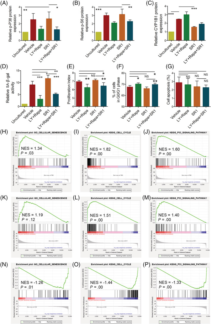
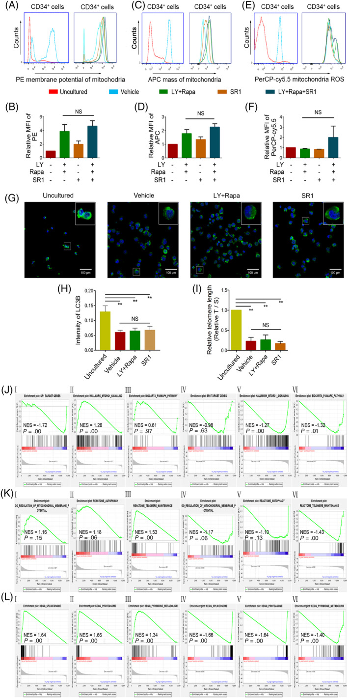
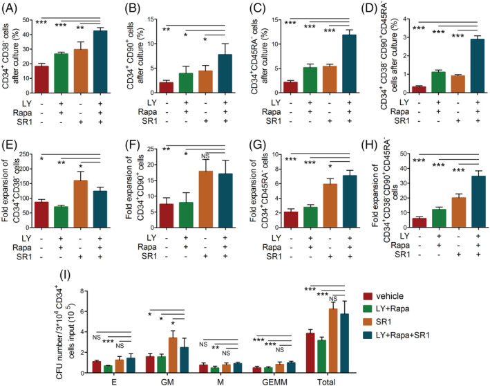
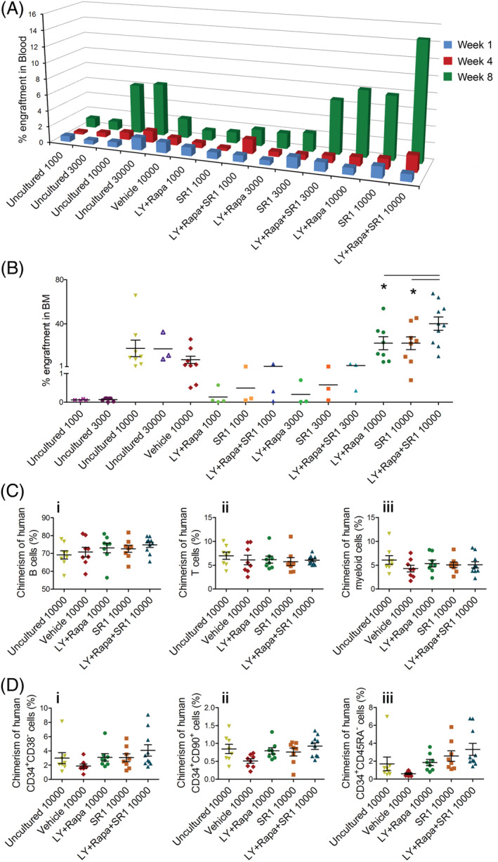
The stemness of ex vivo expanded hematopoietic stem cells (HSCs) is usually compromised by current methods. To explore the failure mechanism of stemness maintenance of human HSCs, which were expanded from human umbilical cord blood (hUCB) CD34 cells, by differentiation inhibitor Stem Regenin 1 (SR1), an antagonist of aryl hydrocarbon receptor, we investigated the activity of p38 mitogen-activated protein kinase α (p38 MAPKα, p38α) and mammalian target of rapamycin complex 1 (mTORC1), and their effect on SR1-expanded hUCB CD34 cells. Our results showed that cellular senescence occurred in the SR1-expanded hUCB CD34 cells in which p38α and mTORC1 were successively activated. Furthermore, their coinhibition resulted in a further decrease in hUCB CD34 cell senescence without an effect on apoptosis, promoted the maintenance of expanded phenotypic HSCs without differentiation inhibition, increased the hematopoietic reconstitution ability of multiple lineages, and potentiated the long-term self-renewal capability of HSCs from SR1-expanded hUCB CD34 cells in NOD/Shi-scid/IL-2Rγ mice. Our mechanistic study revealed that senescence inhibition by our strategy was mainly attributed to downregulation of the splicesome, proteasome formation, and pyrimidine metabolism signaling pathways. These results suggest that coinhibition of activated p38α and mTORC1 potentiates stemness maintenance of HSCs from SR1-expanded hUCB CD34 cells via senescence inhibition. Thus, we established a new strategy to maintain the stemness of ex vivo differentiation inhibitor-expanded human HSCs via coinhibition of multiple independent senescence initiating signal pathways. This senescence inhibition-induced stemness maintenance of ex vivo expanded HSCs could also have an important role in other HSC expansion systems.

摘要

体外扩增的造血干细胞(HSCs)的干性通常会受到当前方法的影响。为了探索分化抑制剂 Stem Regenin 1(SR1)扩增的人脐血(hUCB)CD34 细胞来源的人 HSCs 干性维持失败的机制,我们研究了 p38 丝裂原激活蛋白激酶α(p38 MAPKα,p38α)和哺乳动物雷帕霉素靶蛋白复合物 1(mTORC1)的活性,及其对 SR1 扩增的 hUCB CD34 细胞的影响。结果显示,p38α和 mTORC1 相继激活,导致 SR1 扩增的 hUCB CD34 细胞发生细胞衰老。此外,它们的联合抑制导致 hUCB CD34 细胞衰老进一步减少,而对细胞凋亡没有影响,促进了无分化抑制的扩增表型 HSCs 的维持,增加了多系造血重建能力,并增强了 NOD/Shi-scid/IL-2Rγ小鼠中 SR1 扩增的 hUCB CD34 细胞来源的 HSCs 的长期自我更新能力。我们的机制研究表明,我们的策略通过抑制衰老来增强干性的主要原因是下调剪接体、蛋白酶体形成和嘧啶代谢信号通路。这些结果表明,通过抑制衰老,联合抑制激活的 p38α和 mTORC1 增强了 SR1 扩增的 hUCB CD34 细胞来源的 HSCs 的干性维持。因此,我们建立了一种通过联合抑制多个独立的衰老起始信号通路来维持体外分化抑制剂扩增的人类 HSCs 干性的新策略。这种抑制衰老诱导的体外扩增 HSCs 的干性维持也可能在其他 HSC 扩增系统中发挥重要作用。

https://cdn.ncbi.nlm.nih.gov/pmc/blobs/ea63/7695631/581ddd9ff8a2/SCT3-9-1604-g005.jpg
https://cdn.ncbi.nlm.nih.gov/pmc/blobs/ea63/7695631/ce38d6241fbd/SCT3-9-1604-g001.jpg
https://cdn.ncbi.nlm.nih.gov/pmc/blobs/ea63/7695631/dba051cd2815/SCT3-9-1604-g002.jpg
https://cdn.ncbi.nlm.nih.gov/pmc/blobs/ea63/7695631/85a1004b0929/SCT3-9-1604-g003.jpg
https://cdn.ncbi.nlm.nih.gov/pmc/blobs/ea63/7695631/649ccdd9be15/SCT3-9-1604-g004.jpg
https://cdn.ncbi.nlm.nih.gov/pmc/blobs/ea63/7695631/581ddd9ff8a2/SCT3-9-1604-g005.jpg

相似文献

1
Coinhibition of activated p38 MAPKα and mTORC1 potentiates stemness maintenance of HSCs from SR1-expanded human cord blood CD34 cells via inhibition of senescence.通过抑制衰老,激活的 p38 MAPKα 和 mTORC1 的共抑制增强了 SR1 扩增的人脐血 CD34 细胞中 HSCs 的干性维持。
Stem Cells Transl Med. 2020 Dec;9(12):1604-1616. doi: 10.1002/sctm.20-0129. Epub 2020 Jun 29.
2
Inhibition of p38 MAPK activity promotes ex vivo expansion of human cord blood hematopoietic stem cells.抑制 p38 MAPK 活性可促进人脐血造血干细胞的体外扩增。
Ann Hematol. 2012 Jun;91(6):813-23. doi: 10.1007/s00277-011-1397-7. Epub 2012 Jan 19.
3
Rapamycin enhances long-term hematopoietic reconstitution of ex vivo expanded mouse hematopoietic stem cells by inhibiting senescence.雷帕霉素通过抑制衰老增强体外扩增的小鼠造血干细胞的长期造血重建。
Transplantation. 2014 Jan 15;97(1):20-9. doi: 10.1097/TP.0b013e3182a7fcf8.
4
Low oxygen tension favored expansion and hematopoietic reconstitution of CD34(+) CD38(-) cells expanded from human cord blood-derived CD34(+) Cells.低氧张力有利于从人脐带血来源的CD34(+)细胞扩增而来的CD34(+) CD38(-)细胞的扩增和造血重建。
Biotechnol J. 2016 Jul;11(7):945-53. doi: 10.1002/biot.201500497. Epub 2016 Apr 29.
5
Inhibition of p38 mitogen-activated protein kinase promotes ex vivo hematopoietic stem cell expansion.p38 丝裂原活化蛋白激酶的抑制促进了造血干细胞的体外扩增。
Stem Cells Dev. 2011 Jul;20(7):1143-52. doi: 10.1089/scd.2010.0413. Epub 2011 Feb 24.
6
Differential ability of MSCs isolated from placenta and cord as feeders for supporting ex vivo expansion of umbilical cord blood derived CD34(+) cells.从胎盘和脐带分离的间充质干细胞作为饲养层支持脐血来源的CD34(+)细胞体外扩增的差异能力。
Stem Cell Res Ther. 2015 Oct 19;6:201. doi: 10.1186/s13287-015-0194-y.
7
Safety and efficacy of ex vivo expanded CD34 stem cells in murine and primate models.体外扩增 CD34 干细胞在鼠类和灵长类动物模型中的安全性和有效性。
Stem Cell Res Ther. 2019 Jun 13;10(1):173. doi: 10.1186/s13287-019-1275-0.
8
Generation and function of progenitor T cells from StemRegenin-1-expanded CD34+ human hematopoietic progenitor cells.从 StemRegenin-1 扩增的 CD34+ 人造血祖细胞中产生和功能祖 T 细胞。
Blood Adv. 2019 Oct 22;3(20):2934-2948. doi: 10.1182/bloodadvances.2018026575.
9
Stem cell factor is essential for preserving NOD/SCID reconstitution capacity of ex vivo expanded cord blood CD34(+) cells.干细胞因子对于维持体外扩增的脐血CD34(+)细胞的NOD/SCID重建能力至关重要。
Cell Prolif. 2015 Jun;48(3):293-300. doi: 10.1111/cpr.12186. Epub 2015 Apr 21.
10
Inhibition of MEK/ERK signalling pathway promotes erythroid differentiation and reduces HSCs engraftment in ex vivo expanded haematopoietic stem cells.抑制 MEK/ERK 信号通路可促进红系分化,并减少体外扩增造血干细胞中 HSCs 的植入。
J Cell Mol Med. 2018 Mar;22(3):1464-1474. doi: 10.1111/jcmm.13379. Epub 2017 Oct 10.

引用本文的文献

1
A p38 MAPK-ROS axis fuels proliferation stress and DNA damage during CRISPR-Cas9 gene editing in hematopoietic stem and progenitor cells.p38 MAPK-ROS 轴在造血干细胞和祖细胞中的 CRISPR-Cas9 基因编辑过程中促进增殖应激和 DNA 损伤。
Cell Rep Med. 2024 Nov 19;5(11):101823. doi: 10.1016/j.xcrm.2024.101823. Epub 2024 Nov 12.
2
Time-series analysis of hematopoietic stem cells.造血干细胞的时间序列分析。
Medicine (Baltimore). 2024 Feb 23;103(8):e36509. doi: 10.1097/MD.0000000000036509.
3
Human serum albumin promotes self-renewal and expansion of umbilical cord blood CD34 hematopoietic stem/progenitor cells.

本文引用的文献

1
Long-term ex vivo haematopoietic-stem-cell expansion allows nonconditioned transplantation.长期的体外造血干细胞扩增可实现非条件性移植。
Nature. 2019 Jul;571(7763):117-121. doi: 10.1038/s41586-019-1244-x. Epub 2019 May 29.
2
Pathway enrichment analysis and visualization of omics data using g:Profiler, GSEA, Cytoscape and EnrichmentMap.使用 g:Profiler、GSEA、Cytoscape 和 EnrichmentMap 进行组学数据的通路富集分析和可视化。
Nat Protoc. 2019 Feb;14(2):482-517. doi: 10.1038/s41596-018-0103-9.
3
Hallmarks of Cellular Senescence.
人血清白蛋白促进脐带血CD34造血干/祖细胞的自我更新和扩增。
Ann Transl Med. 2023 Jan 31;11(2):62. doi: 10.21037/atm-22-6383. Epub 2023 Jan 13.
4
Clinical Progress and Preclinical Insights Into Umbilical Cord Blood Transplantation Improvement.脐带血移植改进的临床进展和临床前见解。
Stem Cells Transl Med. 2022 Sep 21;11(9):912-926. doi: 10.1093/stcltm/szac056.
5
DNA barcode to trace the development and differentiation of cord blood stem cells (Review).DNA 条形码追踪脐血干细胞的发育和分化(综述)。
Mol Med Rep. 2021 Dec;24(6). doi: 10.3892/mmr.2021.12489. Epub 2021 Oct 13.
细胞衰老的特征。
Trends Cell Biol. 2018 Jun;28(6):436-453. doi: 10.1016/j.tcb.2018.02.001. Epub 2018 Feb 21.
4
Antagonism of PPAR-γ signaling expands human hematopoietic stem and progenitor cells by enhancing glycolysis.PPAR-γ 信号拮抗作用通过增强糖酵解来扩增人类造血干/祖细胞。
Nat Med. 2018 Mar;24(3):360-367. doi: 10.1038/nm.4477. Epub 2018 Jan 29.
5
AMPK Maintains Cellular Metabolic Homeostasis through Regulation of Mitochondrial Reactive Oxygen Species.AMPK 通过调节线粒体活性氧来维持细胞代谢稳态。
Cell Rep. 2017 Oct 3;21(1):1-9. doi: 10.1016/j.celrep.2017.09.026.
6
SnapShot: Cellular Senescence Pathways.快照:细胞衰老途径。
Cell. 2017 Aug 10;170(4):816-816.e1. doi: 10.1016/j.cell.2017.07.049.
7
Senescence in Health and Disease.健康与疾病中的衰老
Cell. 2017 Jun 1;169(6):1000-1011. doi: 10.1016/j.cell.2017.05.015.
8
Autophagy maintains the metabolism and function of young and old stem cells.自噬维持年轻和衰老干细胞的新陈代谢及功能。
Nature. 2017 Mar 9;543(7644):205-210. doi: 10.1038/nature21388. Epub 2017 Mar 1.
9
Rejuvenating Strategies for Stem Cell-Based Therapies in Aging.衰老过程中基于干细胞疗法的年轻化策略
Cell Stem Cell. 2017 Feb 2;20(2):161-175. doi: 10.1016/j.stem.2017.01.008.
10
Telomere Shortening, Regenerative Capacity, and Cardiovascular Outcomes.端粒缩短、再生能力与心血管结局。
Circ Res. 2017 Mar 31;120(7):1130-1138. doi: 10.1161/CIRCRESAHA.116.309421. Epub 2016 Dec 12.