Department of Neurosurgery, Leiden University Medical Center, Leiden, The Netherlands.
Walaeus Library, Leiden University Medical Center, Leiden, The Netherlands.
Acta Ophthalmol. 2021 Feb;99(1):26-36. doi: 10.1111/aos.14517. Epub 2020 Jun 29.
The effectiveness and safety of surgery for spheno-orbital meningiomas remains subject of debate, as studies often describe different surgical approaches and reconstruction techniques with very heterogeneous outcomes. We aimed to systematically summarize and analyse the literature on spheno-orbital meningiomas regarding presenting symptoms, surgical techniques, outcomes and complications.
Studies were retrieved from eight databases. Original articles were included if in ≥5 patients presenting symptoms, surgical treatment and outcomes were described. Fixed- and random-effects meta-analysis was performed to estimate weighted percentages with 95%CIs of presenting symptoms, outcomes and complications.
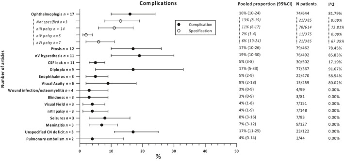
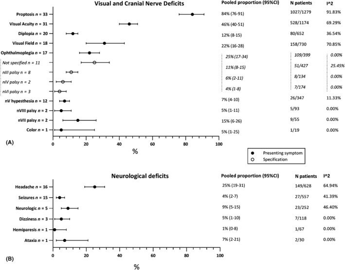
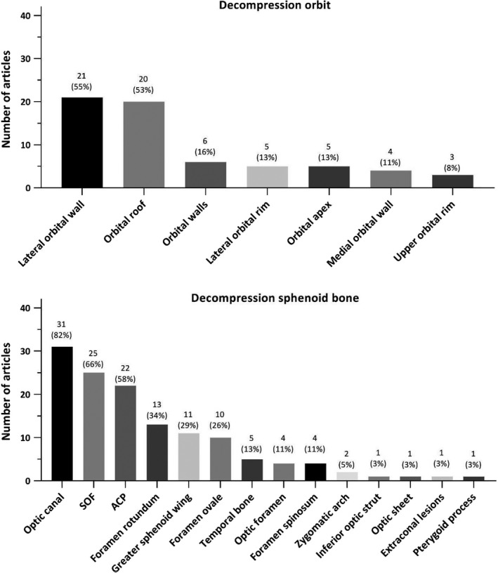
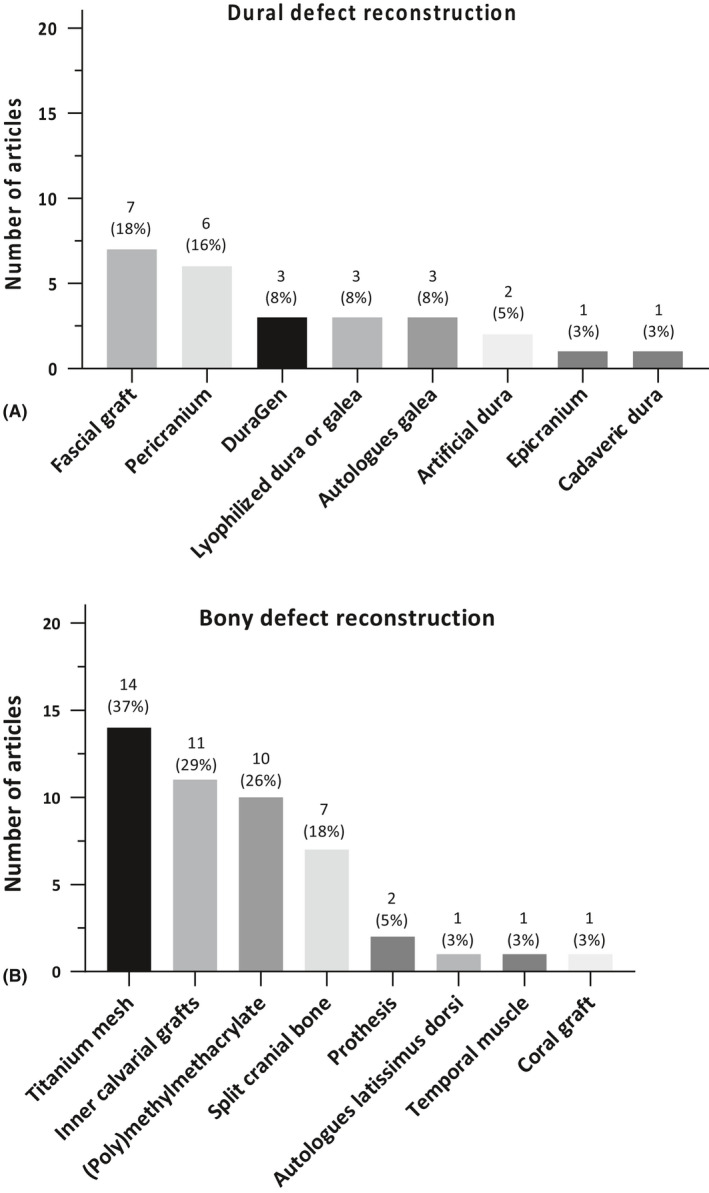
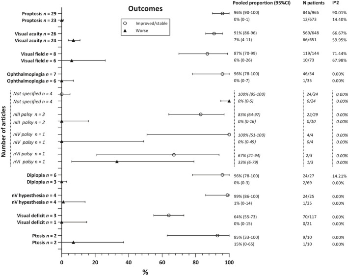
Thirty-eight articles were included describing 1486 patients. Proptosis was the most common presenting symptom (84%; 95%CI 76-91%), followed by unilateral visual acuity deficits (46%; 95%CI 40-51%) and visual field deficits (31%; 95%CI 20-43%). In 35/38 studies (92%), a pterional craniotomy was used. Decompression of the optic canal (82%) and the superior orbital fissure (66%) was most often performed, and usually dural (47%) and bony defects (76%) were reconstructed. In almost all patients, visual acuity (91%; 95%CI 86-96%), visual fields (87%; 95%CI 70-99%) and proptosis (96%; 95%CI 90-100%) improved. Furthermore, surgery showed improvement in 96% (95%CI 78-100%) for both diplopia and ophthalmoplegia. The most common surgical complications were hypesthesia (19%; 95%CI 10-30%), ptosis and diplopia (both 17%; 95%CI, respectively, 10-26% and 5-33%) and ophthalmoplegia (16%; 95%CI 10-24).
Patients with spheno-orbital meningioma usually present with proptosis or unilateral decreased visual acuity. Surgery shows to be effective in improving visual acuity and visual field deficits with mostly minor and well-tolerated complications.
对于蝶眶脑膜瘤的手术治疗效果和安全性仍存在争议,因为研究往往描述了不同的手术入路和重建技术,其结果也存在很大差异。我们旨在系统地总结和分析有关蝶眶脑膜瘤的文献,内容涉及临床表现、手术技术、结果和并发症。
从八个数据库中检索研究。如果≥5 名患者的临床表现、手术治疗和结果得到描述,则纳入原始文章。采用固定效应和随机效应荟萃分析来估计有症状、结局和并发症的加权百分比及其 95%CI。
共纳入 38 篇文章,描述了 1486 例患者。其中,眼球突出是最常见的临床表现(84%;95%CI 76-91%),其次是单侧视力减退(46%;95%CI 40-51%)和视野缺损(31%;95%CI 20-43%)。在 38 项研究中的 35 项(92%)中,使用了翼点入路。最常进行视神经管减压术(82%)和眶上裂减压术(66%),通常进行硬脑膜(47%)和骨缺损重建(76%)。几乎所有患者的视力(91%;95%CI 86-96%)、视野(87%;95%CI 70-99%)和眼球突出度(96%;95%CI 90-100%)均有改善。此外,手术改善了 96%(95%CI 78-100%)的复视和眼肌麻痹症状。最常见的手术并发症为感觉迟钝(19%;95%CI 10-30%)、上睑下垂和复视(各 17%;95%CI 分别为 10-26%和 5-33%)和眼肌麻痹(16%;95%CI 10-24%)。
蝶眶脑膜瘤患者通常表现为眼球突出或单侧视力减退。手术在改善视力和视野缺损方面具有较好的效果,且并发症多为轻微且可耐受。