Planktology, Institute for Chemistry and Biology of the Marine Environment, Carl von Ossietzky University Oldenburg, Carl-von-Ossietzky-Straße 9-11, 26111, Oldenburg, Germany.
Planktology, ICBM, Carl von Ossietzky University Oldenburg, P. O. B. 2503, 26129, Oldenburg, Germany.
Protoplasma. 2020 Nov;257(6):1531-1541. doi: 10.1007/s00709-020-01530-z. Epub 2020 Jul 3.
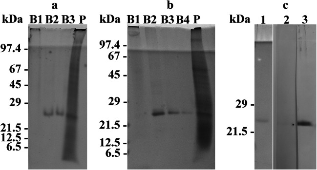
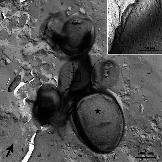
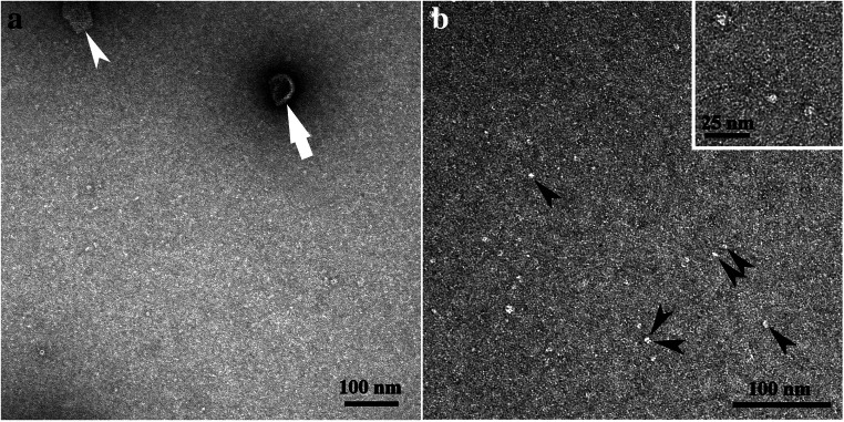
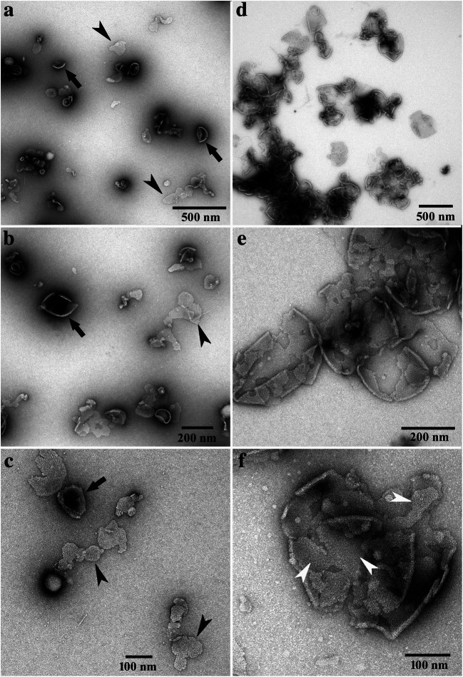
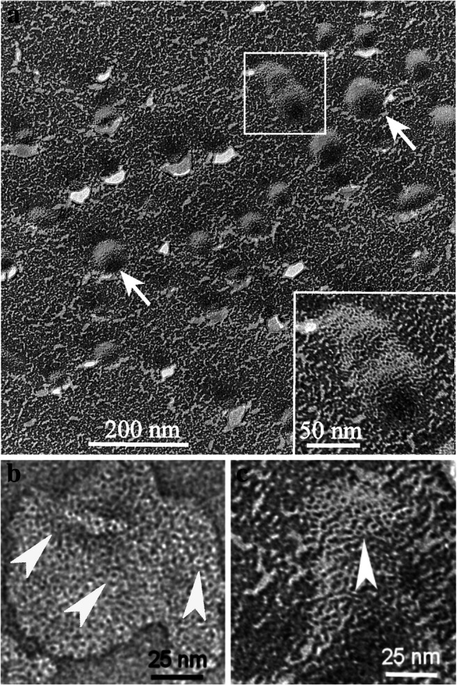
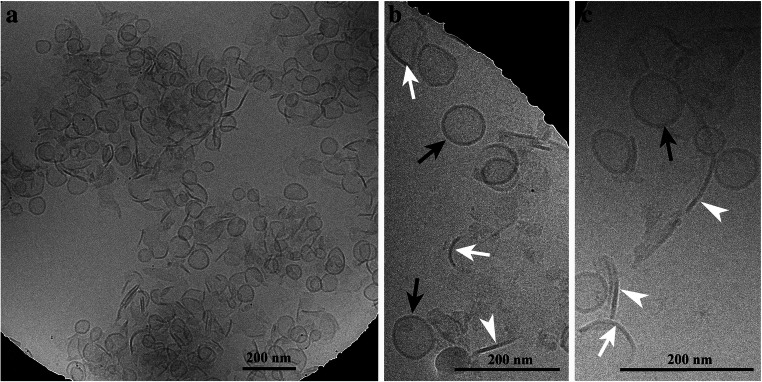
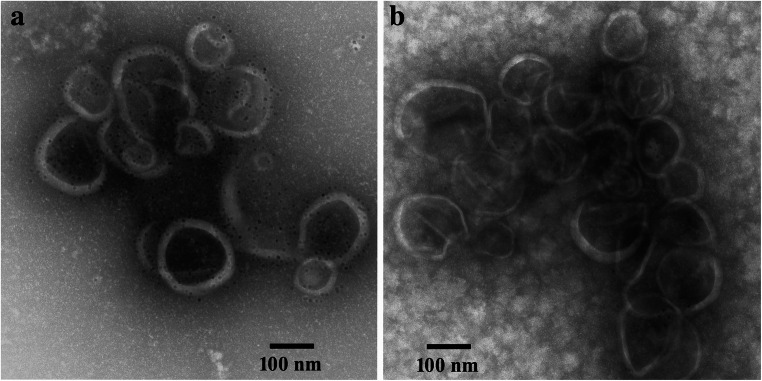
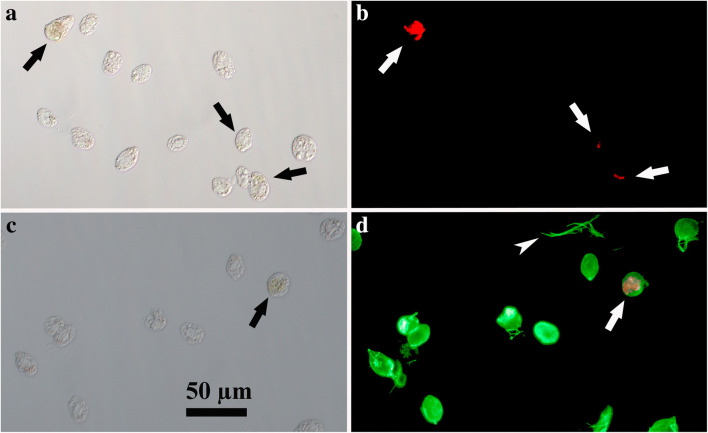
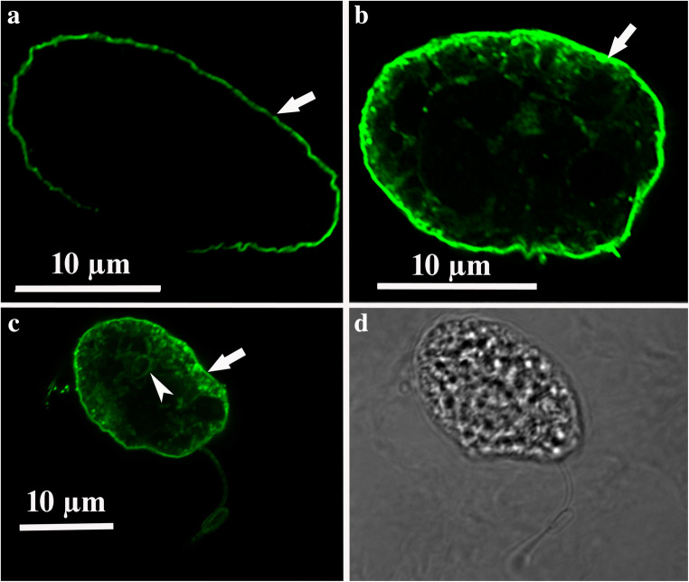
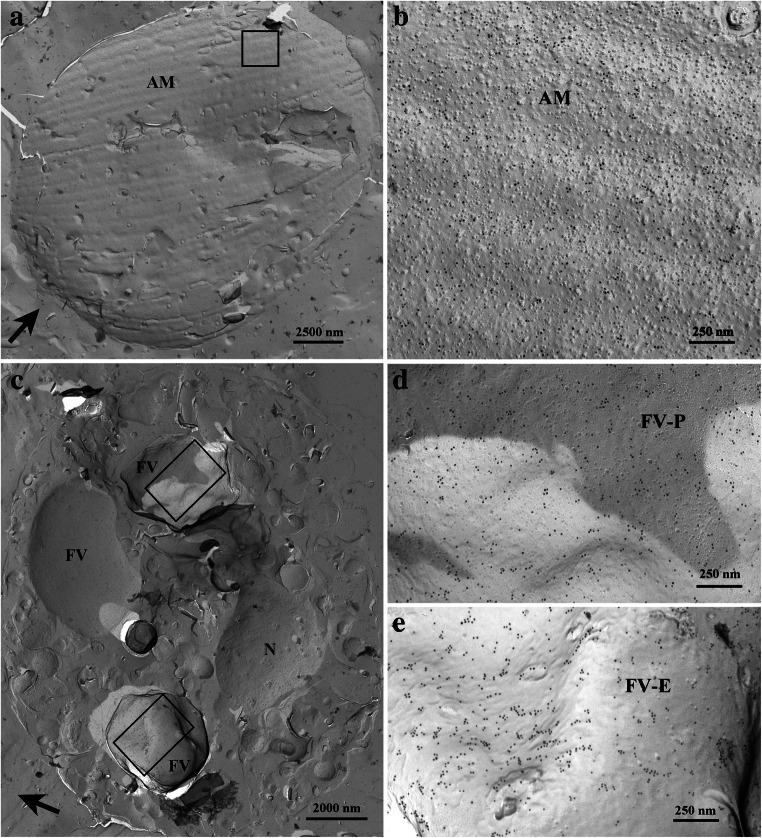
At least 7 proteorhodopsin sequences of Oxyrrhis marina were recently proven in bands obtained by sucrose density gradient centrifugation, and MS analyses revealed that the bands consisted almost of pure, native proteorhodopsins (Rhiel et al. 2020). The proteorhodopsin fractions, i.e., bands B2, B3, and B4 were subjected to transmission electron microscopy. Negative staining revealed that band B2 consisted most likely of monomeric/oligomeric proteorhodopsins with particle dimensions of about 6 nm. Negative staining, freeze-fracture, and cryo-transmission electron microscopy revealed that bands B3 and B4 consisted of vesicular, sheet-like, and cup-shaped structures which all seemed to be composed of protein. Frequently, ring-like protein aggregates were registered at higher magnifications. They measured about 4 nm in diameter with a tiny hole of 1.5 nm in the middle. The bands B2, B3, and B4 were pooled and used to raise an antiserum. Immunoelectron microscopy resulted in intense labeling of the isolated structures. Immunofluorescence light microscopy of formaldehyde-fixed Oxyrrhis cells resulted in intense labeling of the cell periphery. Some cell internal structures became labeled, too. Immunoelectron microscopy of freeze-fractured cells revealed that most likely the membranes of the amphiesmal vesicles were labeled at the cell periphery, while the cell internal label seemed to originate from the food vacuoles.
最近,通过蔗糖密度梯度离心获得的带中证实了至少 7 种 Oxyrrhis marina 的原噬菌视紫质序列,MS 分析表明这些带几乎由纯的天然原噬菌视紫质组成 (Rhiel 等人,2020 年)。原噬菌视紫质部分,即带 B2、B3 和 B4 进行了透射电子显微镜观察。负染色显示带 B2 可能由单体/寡聚原噬菌视紫质组成,颗粒尺寸约为 6nm。负染色、冷冻断裂和 cryo 透射电子显微镜显示带 B3 和 B4 由囊泡状、片状和杯状结构组成,这些结构似乎都由蛋白质组成。在更高的放大倍数下,经常可以看到环状蛋白质聚集体。它们的直径约为 4nm,中间有一个 1.5nm 的小孔。将带 B2、B3 和 B4 混合并用于制备抗血清。免疫电子显微镜导致分离结构的强烈标记。福尔马林固定的 Oxyrrhis 细胞的免疫荧光显微镜观察导致细胞周围的强烈标记。一些细胞内部结构也被标记。冷冻断裂细胞的免疫电子显微镜显示,最有可能的是在细胞周围标记了 Amphiesmal 囊泡的膜,而细胞内部的标记似乎来自食物液泡。