Delia D, Traversari C, Ballinari D, Cattoretti G, Fontanella E, Polli N, Della Porta G
Division of Experimental Oncology A, Istituto Nazionale per lo Studio e la Cura dei Tumori, Milano, Italy.
Immunology. 1988 Aug;64(4):593-8.
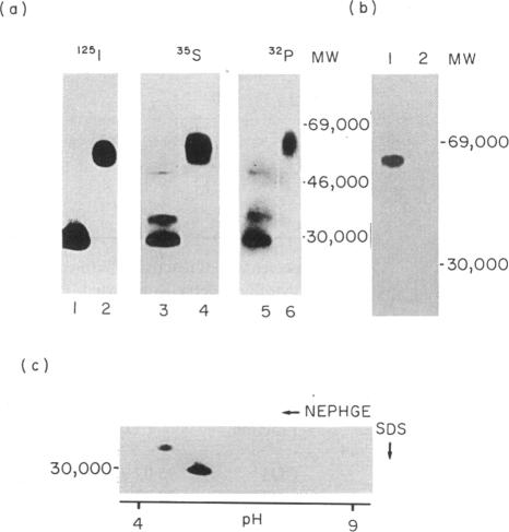
The MLR-3 monoclonal antibody reacts with activated but not with resting lymphocytes. We report that MLR-3 identifies an early activation molecule since its binding is detectable on T cells 1.5-2 hr after in vitro activation. Its expression, therefore, does not require DNA synthesis and precedes, by many hours, that of the receptors for interleukin-2 (IL-2R) and transferrin (TF-R). The MLR-3 antigen is also found on activated thymocytes (including the large early thymic CD3- subset) and B cells. The majority of T- and B-lymphoblastoid cell lines, as well as the myeloid and erythroid cell lines HL60, GM1 and K562, are MLR-3+; conversely, non-haemopoietic cell lines are MLR-3 negative. Seventy percent of B-cell chronic lymphocytic leukaemia and 15% of B non-Hodgkin's lymphomas (B-NHL) are MLR-3+. On tissue sections MLR-3 is reactive with epithelia, sweat glands, hair follicles and Henle's loop but not with vessels, connective, endothelium and many other tissues. In vitro studies show that MLR-3 (1-100 micrograms/ml) significantly alters the thymidine uptake of mitogen-treated lymphocytes:augmentation is found when T and B cells are induced with TPA-Ionomycin and reduction when induced with phytohaemoagglutinin (PHA) or Staphylococcus aureus Cowan strain 1 (SAC), respectively. On SDS-PAGE, MLR-3 immunoprecipitates a disulphide-linked heterodimer of MW 29,000-35,000: both subunits are glycosylated, phosphorylated and exhibit a pI of 4.1 and 5.0, respectively. Our data, particularly the in vitro results, suggest that the MRL-3 molecule could have an important role in the early hours of activation for the progression of resting lymphocytes into mitosis.
MLR-3单克隆抗体与活化淋巴细胞发生反应,而不与静止淋巴细胞反应。我们报告称,MLR-3识别一种早期活化分子,因为在体外活化后1.5 - 2小时即可在T细胞上检测到其结合。因此,其表达不需要DNA合成,且比白细胞介素-2(IL-2R)和转铁蛋白(TF-R)受体的表达提前数小时。在活化的胸腺细胞(包括大型早期胸腺CD3-亚群)和B细胞上也发现了MLR-3抗原。大多数T和B淋巴母细胞系,以及髓系和红系细胞系HL60、GM1和K562均为MLR-3阳性;相反,非造血细胞系为MLR-3阴性。70%的B细胞慢性淋巴细胞白血病和15%的B非霍奇金淋巴瘤(B-NHL)为MLR-3阳性。在组织切片上,MLR-3与上皮、汗腺、毛囊和亨利袢发生反应,但不与血管、结缔组织、内皮及许多其他组织发生反应。体外研究表明,MLR-3(1 - 100微克/毫升)可显著改变丝裂原处理的淋巴细胞的胸腺嘧啶摄取:用佛波酯-离子霉素诱导T和B细胞时摄取增加,而分别用植物血凝素(PHA)或金黄色葡萄球菌考恩1株(SAC)诱导时摄取减少。在SDS-PAGE上,MLR-3免疫沉淀出一个分子量为29,000 - 35,000的二硫键连接的异二聚体:两个亚基均被糖基化、磷酸化,其pI分别为4.1和5.0。我们的数据,尤其是体外实验结果表明,MRL-3分子在静止淋巴细胞进入有丝分裂进程的早期活化阶段可能发挥重要作用。