• 文献检索
  • 文档翻译
  • 深度研究
  • 学术资讯
  • Suppr Zotero 插件Zotero 插件
  • 邀请有礼
  • 套餐&价格
  • 历史记录
应用&插件
Suppr Zotero 插件Zotero 插件浏览器插件Mac 客户端Windows 客户端微信小程序
定价
高级版会员购买积分包购买API积分包
服务
文献检索文档翻译深度研究API 文档MCP 服务
关于我们
关于 Suppr公司介绍联系我们用户协议隐私条款
关注我们

Suppr 超能文献

核心技术专利:CN118964589B侵权必究
粤ICP备2023148730 号-1Suppr @ 2026

文献检索

告别复杂PubMed语法,用中文像聊天一样搜索,搜遍4000万医学文献。AI智能推荐,让科研检索更轻松。

立即免费搜索

文件翻译

保留排版,准确专业,支持PDF/Word/PPT等文件格式,支持 12+语言互译。

免费翻译文档

深度研究

AI帮你快速写综述,25分钟生成高质量综述,智能提取关键信息,辅助科研写作。

立即免费体验

肺动脉高压患者右心室应变-面积环的 5 年预后价值。

5-Year prognostic value of the right ventricular strain-area loop in patients with pulmonary hypertension.

机构信息

Department of Physiology, Research Institute for Health Sciences, Radboud University Medical Center, Philips van Leijdenlaan 15, 6525 EX Nijmegen, The Netherlands.

Department of Cardiology, Research Institute for Health Sciences, Radboud University Medical Center, Geert Grooteplein Zuid 10 6525 GA Nijmegen, The Netherlands.

出版信息

Eur Heart J Cardiovasc Imaging. 2021 Jan 22;22(2):188-195. doi: 10.1093/ehjci/jeaa143.

DOI:10.1093/ehjci/jeaa143
PMID:32632438
原文链接:https://pmc.ncbi.nlm.nih.gov/articles/PMC7822639/
Abstract

AIMS

Patients with pre-capillary pulmonary hypertension (PH) show poor survival, often related to right ventricular (RV) dysfunction. In this study, we assessed the 5-year prognostic value of a novel echocardiographic measure that examines RV function through the temporal relation between RV strain (ϵ) and area (i.e. RV ϵ-area loop) for all-cause mortality in PH patients.

METHODS AND RESULTS

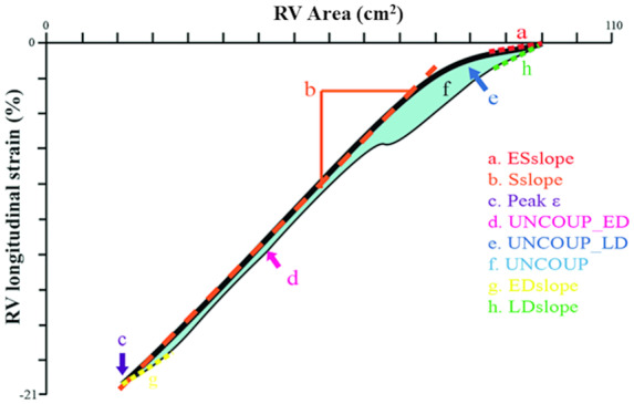
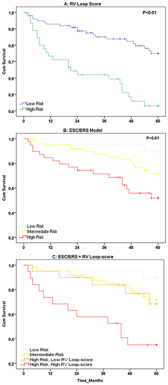
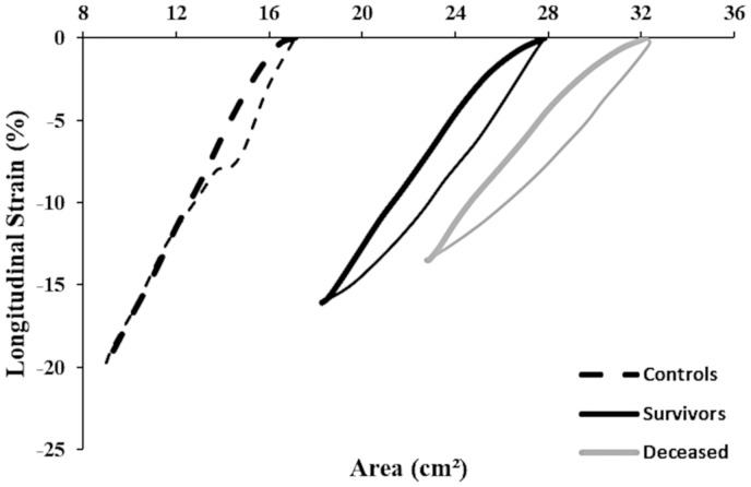
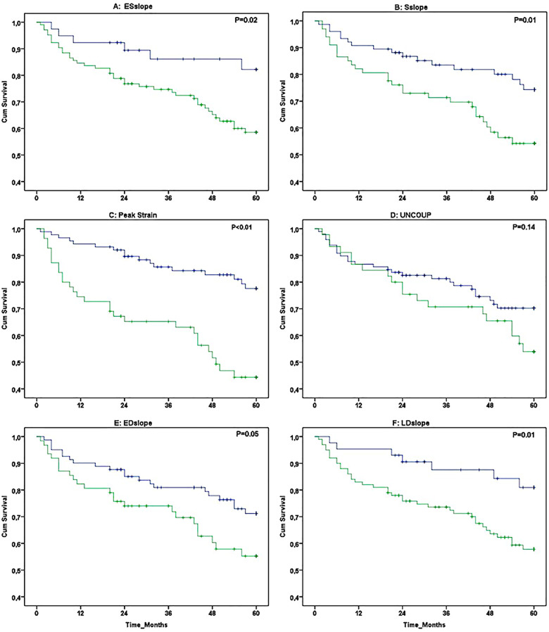
Echocardiographic assessments were performed in 143 PH patients (confirmed by right heart catheterization). Transthoracic echocardiography was utilized to assess RV ϵ-area loop. Using receiver operating characteristic curve-derived cut-off values, we stratified patients in low- vs. high-risk groups for all-cause mortality. Kaplan-Meier survival curves and uni-/multivariable cox-regression models were used to assess RV ϵ-area loop's prognostic value (independent of established predictors: age, sex, N-terminal pro B-type natriuretic peptide, 6-min walking distance). During follow-up 45 (31%) patients died, who demonstrated lower systolic slope, peak ϵ, and late diastolic slope (all P < 0.05) at baseline. Univariate cox-regression analyses identified early systolic slope, systolic slope, peak ϵ, early diastolic uncoupling, and early/late diastolic slope to predict all-cause mortality (all P < 0.05), whilst peak ϵ possessed independent prognostic value (P < 0.05). High RV loop-score (i.e. based on number of abnormal characteristics) showed poorer survival compared to low RV loop-score (Kaplan-Meier: P < 0.01). RV loop-score improved risk stratification in high-risk patients when added to established predictors.

CONCLUSION

Our data demonstrate the potential for RV ϵ-area loops to independently predict all-cause mortality in patients with pre-capillary PH. The non-invasive nature and simplicity of measuring the RV ϵ-area loop, support the potential clinical relevance of (repeated) echocardiography assessment of PH patients.

摘要

目的

患有毛细血管前肺高血压(PH)的患者生存率较差,这通常与右心室(RV)功能障碍有关。在这项研究中,我们评估了一种新的超声心动图指标的 5 年预后价值,该指标通过 RV 应变(ϵ)与面积之间的时间关系(即 RV ϵ-面积环)来评估 PH 患者的全因死亡率。

方法和结果

对 143 例 PH 患者(通过右心导管检查确诊)进行了超声心动图评估。经胸超声心动图用于评估 RV ϵ-面积环。使用接受者操作特征曲线得出的截断值,我们将患者分为低危和高危全因死亡率组。使用 Kaplan-Meier 生存曲线和单变量/多变量 Cox 回归模型评估 RV ϵ-面积环的预后价值(独立于既定预测因子:年龄、性别、N 末端 B 型利钠肽前体、6 分钟步行距离)。在随访期间,有 45 名(31%)患者死亡,他们在基线时表现出较低的收缩斜率、峰值 ϵ 和舒张晚期斜率(均 P<0.05)。单变量 Cox 回归分析确定了早期收缩斜率、收缩斜率、峰值 ϵ、早期舒张去耦和早期/晚期舒张斜率来预测全因死亡率(均 P<0.05),而峰值 ϵ 具有独立的预后价值(P<0.05)。高 RV 环评分(即基于异常特征的数量)与低 RV 环评分相比,生存率较差(Kaplan-Meier:P<0.01)。当 RV 环评分添加到既定预测因子中时,可改善高危患者的风险分层。

结论

我们的数据表明,RV ϵ-面积环可独立预测毛细血管前 PH 患者的全因死亡率。RV ϵ-面积环的非侵入性和测量的简单性支持对 PH 患者进行(重复)超声心动图评估的潜在临床相关性。

https://cdn.ncbi.nlm.nih.gov/pmc/blobs/ec0a/7822639/4efeb732bc0e/jeaa143f4.jpg
https://cdn.ncbi.nlm.nih.gov/pmc/blobs/ec0a/7822639/15c2c31dc8bf/jeaa143f1.jpg
https://cdn.ncbi.nlm.nih.gov/pmc/blobs/ec0a/7822639/ee65245114e0/jeaa143f2.jpg
https://cdn.ncbi.nlm.nih.gov/pmc/blobs/ec0a/7822639/a709e47e34f1/jeaa143f3.jpg
https://cdn.ncbi.nlm.nih.gov/pmc/blobs/ec0a/7822639/4efeb732bc0e/jeaa143f4.jpg

相似文献

1
5-Year prognostic value of the right ventricular strain-area loop in patients with pulmonary hypertension.肺动脉高压患者右心室应变-面积环的 5 年预后价值。
Eur Heart J Cardiovasc Imaging. 2021 Jan 22;22(2):188-195. doi: 10.1093/ehjci/jeaa143.
2
Prognostic value of right ventricular free wall strain in pulmonary hypertension patients with pseudo-normalized tricuspid annular plane systolic excursion values.三尖瓣环平面收缩期位移值假性正常化的肺动脉高压患者右心室游离壁应变的预后价值
Int J Cardiovasc Imaging. 2016 Jun;32(6):905-12. doi: 10.1007/s10554-016-0862-8. Epub 2016 Mar 1.
3
Two-dimensional speckle tracking echocardiography assessed right ventricular function and exercise capacity in pre-capillary pulmonary hypertension.二维斑点追踪超声心动图评估毛细血管前性肺动脉高压患者的右心室功能和运动能力。
Int J Cardiovasc Imaging. 2019 Aug;35(8):1499-1508. doi: 10.1007/s10554-019-01605-w. Epub 2019 Apr 29.
4
Outcome prediction by quantitative right ventricular function assessment in 575 subjects evaluated for pulmonary hypertension.575 例肺动脉高压评估患者的定量右心功能评估的预后预测。
Circ Cardiovasc Imaging. 2013 Sep;6(5):711-21. doi: 10.1161/CIRCIMAGING.113.000640. Epub 2013 Jun 27.
5
Comprehensive assessment of right ventricular function in patients with pulmonary hypertension with global longitudinal peak systolic strain derived from multiple right ventricular views.利用多个右心室视图得出的整体纵向收缩期峰值应变对肺动脉高压患者的右心室功能进行综合评估。
J Am Soc Echocardiogr. 2014 Jun;27(6):657-665.e3. doi: 10.1016/j.echo.2014.02.001. Epub 2014 Mar 20.
6
Clinical Determinants and Prognostic Implications of Right Ventricular Dysfunction in Pulmonary Hypertension Caused by Chronic Lung Disease.慢性肺部疾病引起的肺动脉高压患者右心功能障碍的临床决定因素和预后意义。
J Am Heart Assoc. 2019 Jan 22;8(2):e011464. doi: 10.1161/JAHA.118.011464.
7
Cardiac Magnetic Resonance Imaging-Based Right Ventricular Strain Analysis for Assessment of Coupling and Diastolic Function in Pulmonary Hypertension.基于心脏磁共振成像的右心室应变分析评估肺动脉高压的耦联和舒张功能。
JACC Cardiovasc Imaging. 2019 Nov;12(11 Pt 1):2155-2164. doi: 10.1016/j.jcmg.2018.12.032. Epub 2019 Mar 13.
8
Association of right atrial conduit phase with right ventricular lusitropic function in pulmonary hypertension.右心房导联相位与肺动脉高压右心室舒缩功能的关系。
Int J Cardiovasc Imaging. 2020 Apr;36(4):633-642. doi: 10.1007/s10554-019-01763-x. Epub 2020 Jan 8.
9
Three-dimensional echocardiography and 2D-3D speckle-tracking imaging in chronic pulmonary hypertension: diagnostic accuracy in detecting hemodynamic signs of right ventricular (RV) failure.三维超声心动图和二维-三维斑点追踪成像在慢性肺动脉高压中的应用:检测右心室(RV)衰竭血流动力学征象的诊断准确性
J Am Heart Assoc. 2015 Mar 19;4(3):e001584. doi: 10.1161/JAHA.114.001584.
10
Assessment of right ventricular longitudinal strain by 2D speckle tracking imaging compared with RV function and hemodynamics in pulmonary hypertension.二维斑点追踪成像评估右心室纵向应变与肺动脉高压患者右心室功能及血流动力学的比较
Int J Cardiovasc Imaging. 2017 Nov;33(11):1737-1748. doi: 10.1007/s10554-017-1182-3. Epub 2017 May 29.

引用本文的文献

1
Challenges and opportunities in assessing right ventricular structure and function: a Roadmap for standardization, clinical implementation and research.评估右心室结构和功能的挑战与机遇:标准化、临床应用及研究路线图
Nat Rev Cardiol. 2025 Jun 25. doi: 10.1038/s41569-025-01180-9.
2
Echocardiographic Parameters of the Right Ventricle in Patients With Pulmonary Hypertension: A Review.肺动脉高压患者右心室的超声心动图参数:综述
Korean Circ J. 2025 Apr;55(4):259-274. doi: 10.4070/kcj.2024.0313. Epub 2025 Jan 31.
3
Prognostic values of right ventricular echocardiography functional parameters for mortality prediction in precapillary pulmonary hypertension: a systematic review and meta-analysis.

本文引用的文献

1
Prognostic value of right ventricular longitudinal strain in patients with pulmonary hypertension: a systematic review and meta-analysis.右心室纵向应变对肺动脉高压患者预后的价值:系统评价和荟萃分析。
Eur Heart J Cardiovasc Imaging. 2019 Apr 1;20(4):475-484. doi: 10.1093/ehjci/jey120.
2
Prognostic Value of Right Ventricular Strain Using Speckle-Tracking Echocardiography in Pulmonary Hypertension: A Systematic Review and Meta-analysis.斑点追踪超声心动图右心室应变对肺动脉高压的预后价值:系统评价和荟萃分析。
Can J Cardiol. 2018 Aug;34(8):1069-1078. doi: 10.1016/j.cjca.2018.04.016. Epub 2018 Apr 25.
3
Echocardiographic-Derived Strain-Area Loop of the Right Ventricle is Related to Pulmonary Vascular Resistance in Pulmonary Arterial Hypertension.
右心室超声心动图功能参数对毛细血管前性肺动脉高压患者死亡率预测的预后价值:一项系统评价和荟萃分析
Egypt Heart J. 2024 Aug 14;76(1):105. doi: 10.1186/s43044-024-00539-6.
4
Left ventricular strain-volume loops in bicuspid aortic valve disease: new insights in cardiomechanics.二叶式主动脉瓣疾病中的左心室应变-容积环:心脏力学的新见解
Eur Heart J Imaging Methods Pract. 2024 Mar 15;2(2):qyae020. doi: 10.1093/ehjimp/qyae020. eCollection 2024 Apr.
5
Left and right ventricular strain-volume/area loops: a narrative review of current physiological understanding and potential clinical value.左、右心室应变-容积/面积环:当前生理学认识及潜在临床价值的叙述性综述
Echo Res Pract. 2024 May 21;11(1):12. doi: 10.1186/s44156-024-00046-z.
6
Echocardiographic pressure-strain loop-derived stroke work of the right ventricle: validation against the gold standard.超声心动图压力-应变环衍生的右心室每搏功:与金标准的对比验证
ESC Heart Fail. 2023 Oct;10(5):3209-3215. doi: 10.1002/ehf2.14453. Epub 2023 Jul 6.
7
Assessment of Right Ventricular Function-a State of the Art.右心室功能评估——现状。
Curr Heart Fail Rep. 2023 Jun;20(3):194-207. doi: 10.1007/s11897-023-00600-6. Epub 2023 Jun 5.
8
Association of left ventricular strain-volume loop characteristics with adverse events in patients with heart failure with preserved ejection fraction.左心室应变-容积环特征与射血分数保留心力衰竭患者不良事件的相关性。
Eur Heart J Cardiovasc Imaging. 2023 Aug 23;24(9):1168-1176. doi: 10.1093/ehjci/jead117.
9
Right ventricular volume-strain loops using 3D echocardiography-derived mesh models: proof-of-concept application on patients undergoing different types of open-heart surgery.使用三维超声心动图衍生网格模型的右心室容积-应变环:在接受不同类型心脏直视手术患者中的概念验证应用
Quant Imaging Med Surg. 2022 Jul;12(7):3679-3691. doi: 10.21037/qims-21-1204.
10
Speckle Tracking Imaging in Patients with Pulmonary Hypertension.肺动脉高压患者的斑点追踪成像
J Cardiovasc Imaging. 2021 Jul;29(3):236-251. doi: 10.4250/jcvi.2020.0192. Epub 2021 Mar 18.
超声心动图衍生的右心室应变面积环与肺动脉高压中的肺血管阻力相关。
JACC Cardiovasc Imaging. 2017 Oct;10(10 Pt B):1286-1288. doi: 10.1016/j.jcmg.2017.05.018. Epub 2017 Aug 16.
4
The Giessen Pulmonary Hypertension Registry: Survival in pulmonary hypertension subgroups.吉森肺动脉高压注册研究:肺动脉高压亚组的生存情况。
J Heart Lung Transplant. 2017 Sep;36(9):957-967. doi: 10.1016/j.healun.2017.02.016. Epub 2017 Feb 17.
5
Exploratory assessment of left ventricular strain-volume loops in severe aortic valve diseases.重度主动脉瓣疾病中左心室应变-容积环的探索性评估
J Physiol. 2017 Jun 15;595(12):3961-3971. doi: 10.1113/JP273526. Epub 2017 Mar 9.
6
Left and right ventricular longitudinal strain-volume/area relationships in elite athletes.精英运动员左、右心室纵向应变-容积/面积关系
Int J Cardiovasc Imaging. 2016 Aug;32(8):1199-211. doi: 10.1007/s10554-016-0910-4. Epub 2016 May 21.
7
2015 ESC/ERS Guidelines for the diagnosis and treatment of pulmonary hypertension: The Joint Task Force for the Diagnosis and Treatment of Pulmonary Hypertension of the European Society of Cardiology (ESC) and the European Respiratory Society (ERS): Endorsed by: Association for European Paediatric and Congenital Cardiology (AEPC), International Society for Heart and Lung Transplantation (ISHLT).2015年欧洲心脏病学会(ESC)/欧洲呼吸学会(ERS)肺动脉高压诊断和治疗指南:欧洲心脏病学会(ESC)和欧洲呼吸学会(ERS)肺动脉高压诊断和治疗联合工作组:得到以下组织认可:欧洲儿科和先天性心脏病协会(AEPC)、国际心肺移植学会(ISHLT)。
Eur Heart J. 2016 Jan 1;37(1):67-119. doi: 10.1093/eurheartj/ehv317. Epub 2015 Aug 29.
8
RV Dyssynchrony by Speckle Tracking Strain in Pulmonary Arterial Hypertension: Will This Outcome Variable Take Root?基于斑点追踪应变评估肺动脉高压患者右心室不同步性:这一结局变量会成为研究热点吗?
JACC Cardiovasc Imaging. 2015 Jun;8(6):653-5. doi: 10.1016/j.jcmg.2015.04.003.
9
Abnormal right ventricular relaxation in pulmonary hypertension.肺动脉高压时右心室舒张异常。
Pulm Circ. 2015 Jun;5(2):370-5. doi: 10.1086/681268.
10
Right ventricular architecture responsible for mechanical performance: unifying role of ventricular septum.负责机械性能的右心室结构:室间隔的统一作用。
J Thorac Cardiovasc Surg. 2014 Dec;148(6):3166-71.e1-4. doi: 10.1016/j.jtcvs.2014.05.044. Epub 2014 May 21.