From the Department of Radiology and Biomedical Imaging (L.J.S., L.A.D., I.T.S., J.J.W., J.S., M.D.L., L.A., A.B., J.D., F.H., D.C., J.C.), Department of Internal Medicine, Section of Rheumatology (R.R.M., L.L., R.J.B.), Department of Immunobiology (N.J.), and Department of Pathology (V.P., X.Z.), Yale University School of Medicine, 300 Cedar St, New Haven, CT 06520; Institute of Radiology, Charité-Universitätsmedizin Berlin, corporate member of Freie Universität Berlin, Humboldt-Universität, and Berlin Institute of Health, Berlin, Germany (L.J.S., L.A.D., I.T.S., L.A.); Visage Imaging, San Diego, Calif (M.D.L.); Department of Biomedical Engineering, Yale School of Engineering and Applied Science, New Haven, Conn (J.D.); and Department of Radiology, Hadassah Hebrew University Medical Center, Jerusalem, Israel (S.N.G.).
Radiology. 2020 Sep;296(3):575-583. doi: 10.1148/radiol.2020200373. Epub 2020 Jul 7.
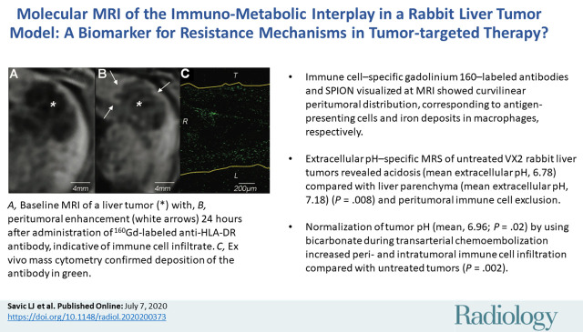
Background The immuno-metabolic interplay has gained interest for determining and targeting immunosuppressive tumor micro-environments that remain a barrier to current immuno-oncologic therapies in hepatocellular carcinoma. Purpose To develop molecular MRI tools to reveal resistance mechanisms to immuno-oncologic therapies caused by the immuno-metabolic interplay in a translational liver cancer model. Materials and Methods A total of 21 VX2 liver tumor-bearing New Zealand white rabbits were used between October 2018 and February 2020. Rabbits were divided into three groups. Group A ( = 3) underwent intra-arterial infusion of gadolinium 160 (Gd)-labeled anti-human leukocyte antigen-DR isotope (HLA-DR) antibodies to detect antigen-presenting immune cells. Group B ( = 3) received rhodamine-conjugated superparamagnetic iron oxide nanoparticles (SPIONs) intravenously to detect macrophages. These six rabbits underwent 3-T MRI, including T1- and T2-weighted imaging, before and 24 hours after contrast material administration. Group C ( = 15) underwent extracellular pH mapping with use of MR spectroscopy. Of those 15 rabbits, six underwent conventional transarterial chemoembolization (TACE), four underwent conventional TACE with extracellular pH-buffering bicarbonate, and five served as untreated controls. MRI signal intensity distribution was validated by using immunohistochemistry staining of HLA-DR and CD11b, Prussian blue iron staining, fluorescence microscopy of rhodamine, and imaging mass cytometry (IMC) of gadolinium. Statistical analysis included Mann-Whitney and Kruskal-Wallis tests. Results T1-weighted MRI with Gd-labeled antibodies revealed localized peritumoral ring enhancement, which corresponded to gadolinium distribution detected with IMC. T2-weighted MRI with SPIONs showed curvilinear signal intensity representing selective peritumoral deposition in macrophages. Extracellular pH-specific MR spectroscopy of untreated liver tumors showed acidosis (mean extracellular pH, 6.78 ± 0.09) compared with liver parenchyma (mean extracellular pH, 7.18 ± 0.03) ( = .008) and peritumoral immune cell exclusion. Normalization of tumor extracellular pH (mean, 6.96 ± 0.05; = .02) using bicarbonate during TACE increased peri- and intratumoral immune cell infiltration ( = .002). Conclusion MRI in a rabbit liver tumor model was used to visualize resistance mechanisms mediated by the immuno-metabolic interplay that inform susceptibility and response to immuno-oncologic therapies, providing a therapeutic strategy to restore immune permissiveness in liver cancer. © RSNA, 2020
背景 免疫代谢相互作用已成为确定和靶向免疫抑制肿瘤微环境的研究热点,而肿瘤微环境正是目前肝癌免疫肿瘤治疗的障碍。目的 开发分子 MRI 工具,以揭示在转化性肝癌模型中由免疫代谢相互作用引起的对免疫肿瘤治疗的抵抗机制。材料与方法 2018 年 10 月至 2020 年 2 月期间,共使用 21 只 VX2 肝癌荷瘤新西兰白兔。将兔子分为三组。A 组(n = 3)经肝动脉内注射钆 160(Gd)标记的抗人白细胞抗原-DR 同位素(HLA-DR)抗体,以检测抗原呈递免疫细胞。B 组(n = 3)经静脉内注射罗丹明标记的超顺磁性氧化铁纳米颗粒(SPIONs),以检测巨噬细胞。这 6 只兔子在注射对比剂前后分别接受了 3T MRI 检查,包括 T1 和 T2 加权成像。C 组(n = 15)采用磁共振波谱法进行细胞外 pH 图谱绘制。其中 15 只兔子中,6 只进行了常规经动脉化疗栓塞(TACE),4 只进行了常规 TACE 联合细胞外 pH 缓冲碳酸氢盐,5 只作为未治疗对照组。采用 HLA-DR 和 CD11b 的免疫组织化学染色、普鲁士蓝铁染色、罗丹明荧光显微镜检查和钆的成像质谱细胞术(IMC)对 MRI 信号强度分布进行验证。统计分析包括 Mann-Whitney 和 Kruskal-Wallis 检验。结果 T1 加权 MRI 联合 Gd 标记抗体显示局部肿瘤周围环形增强,与 IMC 检测到的钆分布相对应。T2 加权 MRI 联合 SPIONs 显示曲线信号强度,代表选择性肿瘤周围巨噬细胞沉积。未经治疗的肝癌肿瘤的细胞外 pH 特异性磁共振波谱显示酸中毒(平均细胞外 pH,6.78 ± 0.09),与肝实质(平均细胞外 pH,7.18 ± 0.03)相比( =.008)和肿瘤周围免疫细胞排斥。TACE 期间使用碳酸氢盐使肿瘤细胞外 pH 正常化(平均 6.96 ± 0.05; =.02)增加了肿瘤周围和肿瘤内免疫细胞浸润( =.002)。结论 在兔肝癌肿瘤模型中进行的 MRI 检查可用于可视化由免疫代谢相互作用介导的抵抗机制,这些抵抗机制可提供有关对免疫肿瘤治疗的敏感性和反应的信息,并提供一种恢复肝癌免疫许可的治疗策略。© RSNA,2020